Study Guides

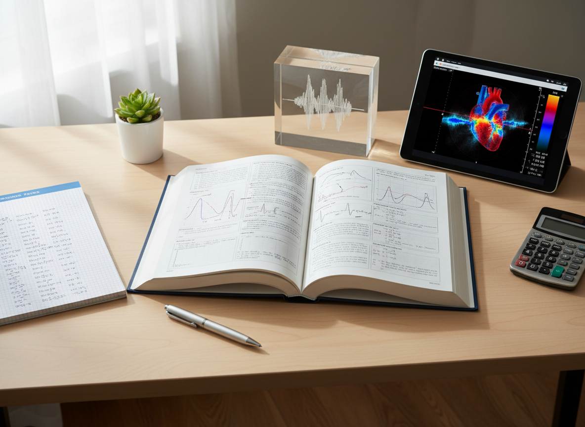
Explore concise introductions and quick-reference tips designed to complement your study sessions and accelerate progress in ultrasound protocol mastery.

A meticulously arranged study desk dedicated to ultrasound physics, featuring an open textbook filled with Doppler waveforms and annotated diagrams, a spiral notebook with neatly written formulas, and a digital tablet showing a color flow image. A small model of a sound wave diagram etched into a transparent acrylic block sits to one side. The desk surface is a light wood grain, with minimal, tidy accessories. Soft daylight from a nearby window washes across the scene, casting gentle shadows and subtle highlights on the pages. Photographic realism, shot from a slightly elevated angle with moderate depth of field, keeping all educational materials sharp. The overall atmosphere is focused, organized, and academically driven, ideal for serious ultrasound students.
A sleek ultrasound machine console with a high-resolution monitor displaying a clear grayscale abdominal scan image, positioned on a clean, reflective hospital floor beside a neatly made examination bed covered with crisp white linens. Color-coded gel bottles and coiled transducer cables rest in organized trays on the machine. Soft, cool overhead clinical lighting creates gentle reflections on the smooth plastic surfaces and subtle shadows underneath the wheels. Photographic realism at eye level with a slight three-quarter angle, capturing the entire system in sharp focus while the background examination room elements fall into a mild blur. The mood is professional, calm, and highly organized, conveying a modern learning environment for ultrasound students.

Ultrasound Basics

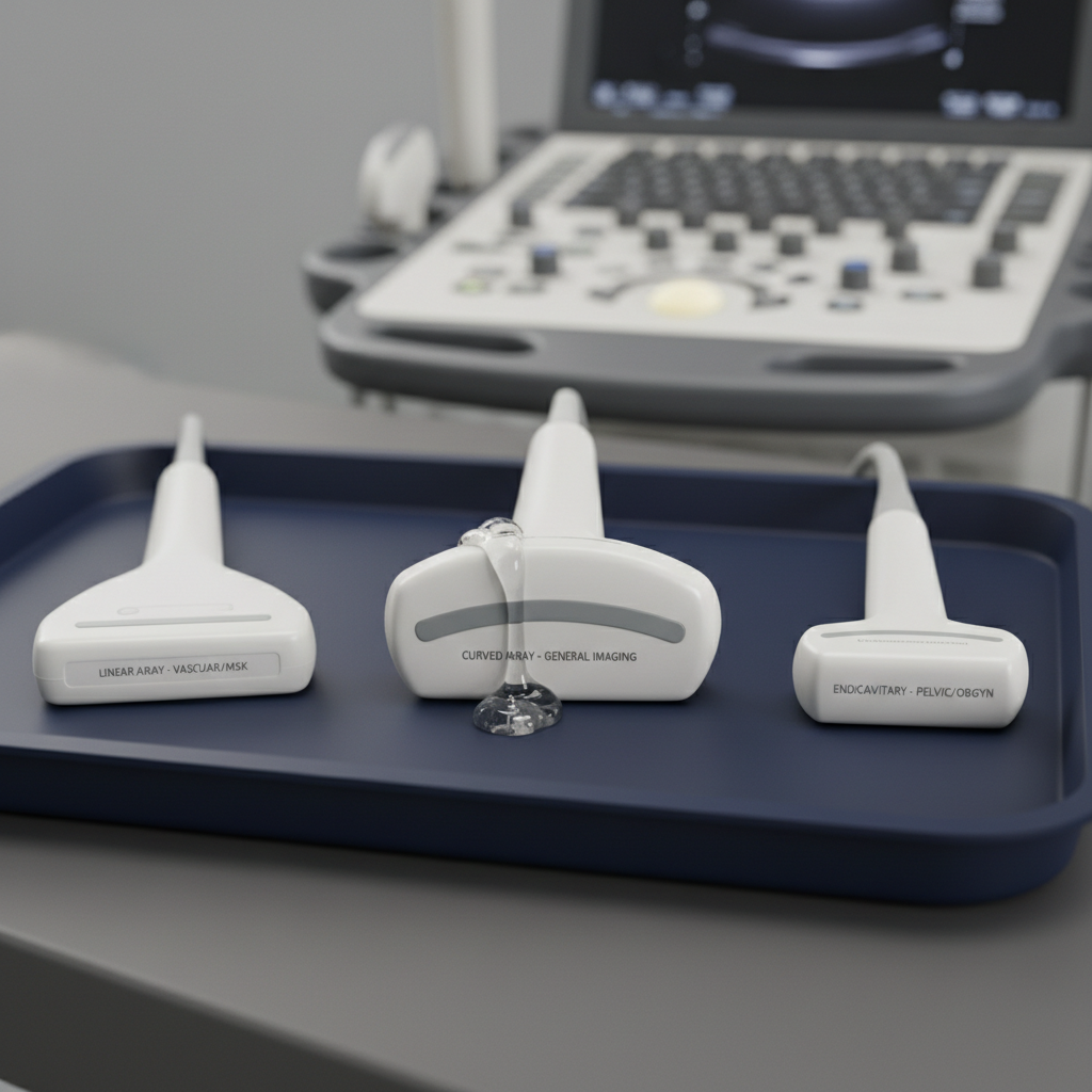
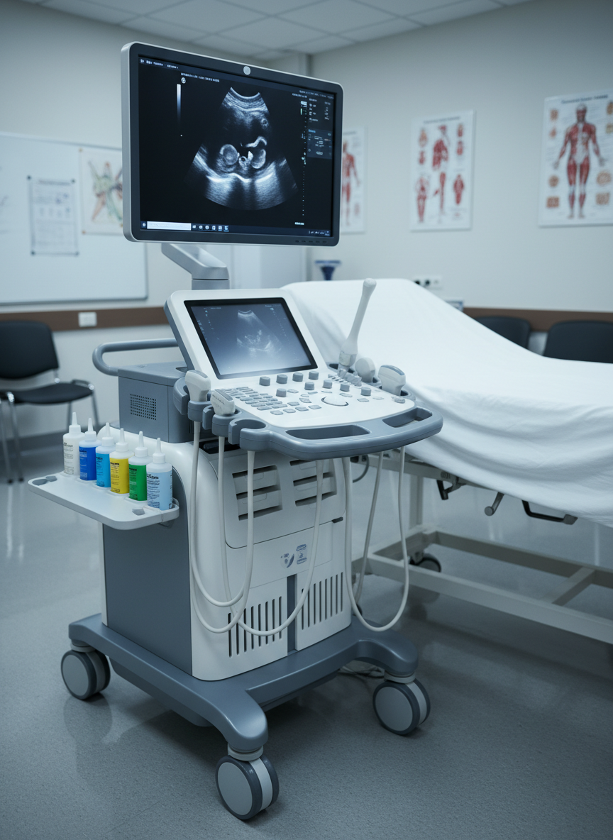
This resource guides you through core ultrasound concepts, physics, and practical study strategies to build confidence and mastery across abdomen, pelvis, and vascular protocols.

Key Concepts

Quick, organized breakdowns help you skim for review or dive deep into topics like physics, anatomy, and protocol steps for abdomen, pelvis, and vessels.

A close-up of three different ultrasound transducers laid out on a matte dark-blue non-slip exam tray: a curved array, linear array, and endocavitary probe, each with finely detailed labels and smooth, slightly glossy white casings. Small droplets of clear gel glisten on one probe surface, showing realistic texture. The tray sits on a neutral gray workstation with a blurred ultrasound console in the background. Soft studio lighting from the upper left creates gentle highlights and crisp but unobtrusive shadows, emphasizing shapes and textures. Photographic realism with a shallow depth of field and a centered composition, creating a focused, educational atmosphere that feels clean, professional, and technical.