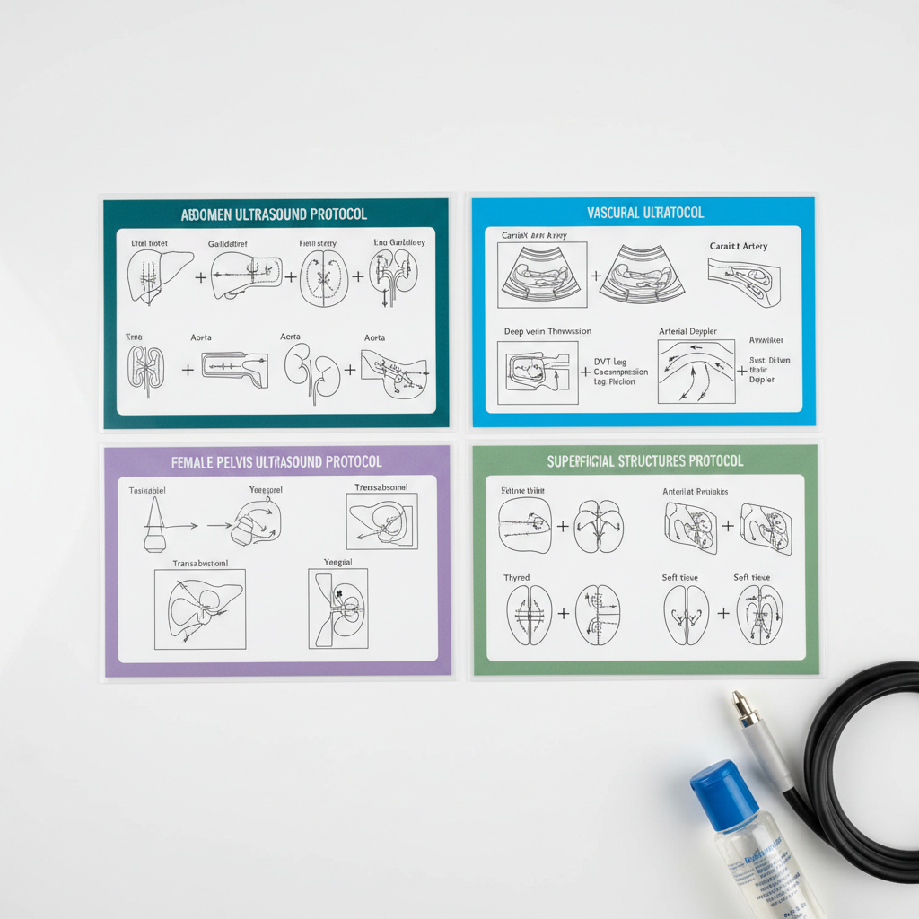
Arterial Venous Protocols

Master core ultrasound approaches for abdomen, superficial, female pelvis, and vascular studies with practical tips, examples, and step-by-step workflows.
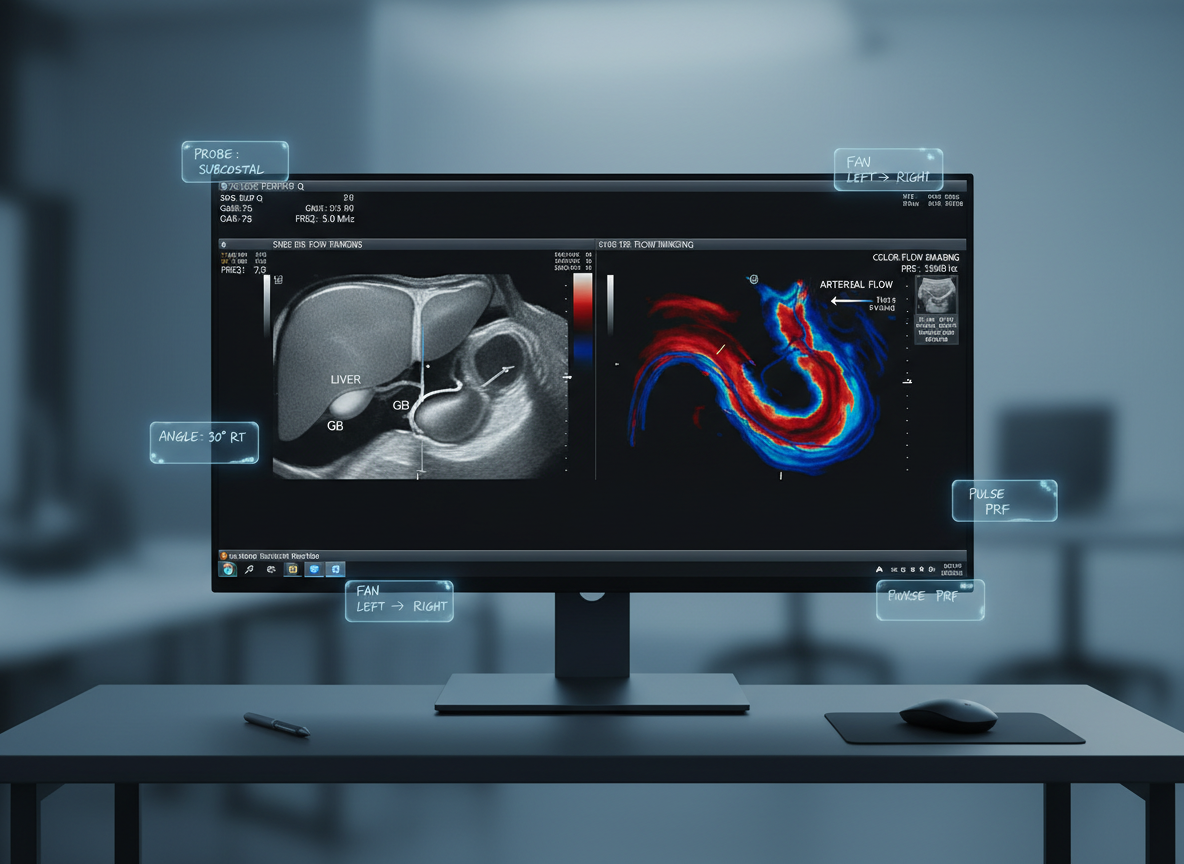
A close-up of three different ultrasound transducers laid out on a matte dark-blue non-slip exam tray: a curved array, linear array, and endocavitary probe, each with finely detailed labels and smooth, slightly glossy white casings. Small droplets of clear gel glisten on one probe surface, showing realistic texture. The tray sits on a neutral gray workstation with a blurred ultrasound console in the background. Soft studio lighting from the upper left creates gentle highlights and crisp but unobtrusive shadows, emphasizing shapes and textures. Photographic realism with a shallow depth of field and a centered composition, creating a focused, educational atmosphere that feels clean, professional, and technical.
A high-resolution monitor displaying a split-screen ultrasound study: on the left, a labeled grayscale image of the liver and gallbladder; on the right, a color Doppler vascular image with vivid reds and blues indicating flow direction. Around the monitor frame, small digital sticky-note style labels show brief study tips like probe positions and angle markers. The monitor rests on a dark, minimal workstation, with the background fading into soft blur. Cool, diffused studio lighting creates a neutral, high-tech ambiance. Photographic realism at a straight-on eye-level composition, with the screen perfectly centered and sharply detailed, conveying a modern, digital study resource for abdominal and vascular ultrasound protocols.

Vascular Protocols

Explore arterial and venous ultrasound protocols, including patient positioning, measurement techniques, and protocol-driven workflows to improve accuracy and efficiency in exams.

An overhead view of a neatly organized set of laminated ultrasound protocol cards laid out on a smooth white tabletop, each card clearly showing step-by-step diagrams for abdomen, superficial structures, female pelvis, and vascular exams. Thin colored borders distinguish each category: deep teal for abdomen, soft purple for pelvis, bright blue for vascular, and muted green for superficial structures. A small, transparent bottle of ultrasound gel and a carefully coiled probe cable appear at the edge of the frame. Soft, even studio lighting eliminates harsh shadows, creating a clean, clinical look. Photographic realism with sharp focus across the entire image and a balanced, symmetrical composition, evoking clarity, structure, and confidence for students mastering complete scanning protocols.
A meticulously arranged study desk dedicated to ultrasound physics, featuring an open textbook filled with Doppler waveforms and annotated diagrams, a spiral notebook with neatly written formulas, and a digital tablet showing a color flow image. A small model of a sound wave diagram etched into a transparent acrylic block sits to one side. The desk surface is a light wood grain, with minimal, tidy accessories. Soft daylight from a nearby window washes across the scene, casting gentle shadows and subtle highlights on the pages. Photographic realism, shot from a slightly elevated angle with moderate depth of field, keeping all educational materials sharp. The overall atmosphere is focused, organized, and academically driven, ideal for serious ultrasound students.
A sleek ultrasound machine console with a high-resolution monitor displaying a clear grayscale abdominal scan image, positioned on a clean, reflective hospital floor beside a neatly made examination bed covered with crisp white linens. Color-coded gel bottles and coiled transducer cables rest in organized trays on the machine. Soft, cool overhead clinical lighting creates gentle reflections on the smooth plastic surfaces and subtle shadows underneath the wheels. Photographic realism at eye level with a slight three-quarter angle, capturing the entire system in sharp focus while the background examination room elements fall into a mild blur. The mood is professional, calm, and highly organized, conveying a modern learning environment for ultrasound students.

Vascular Case Studies