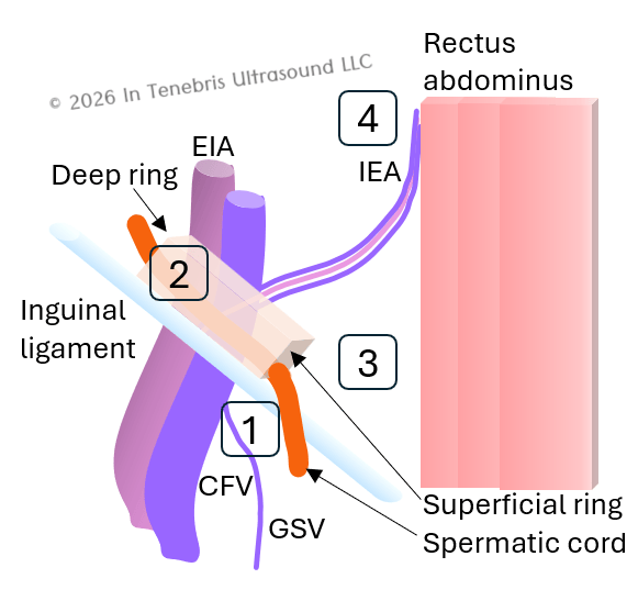
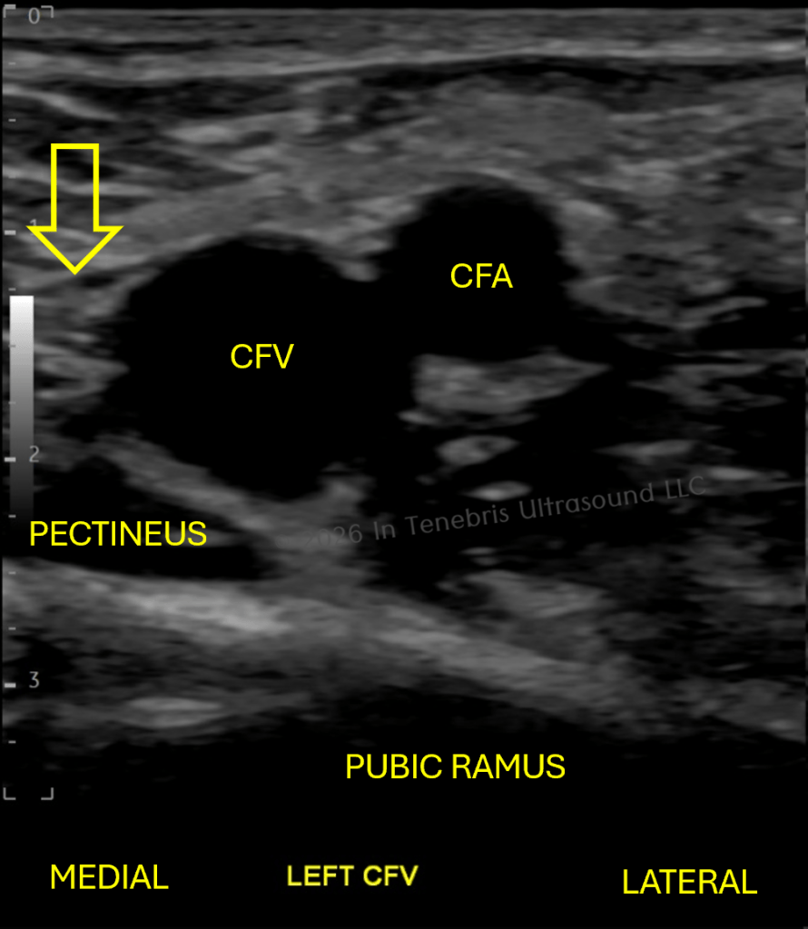
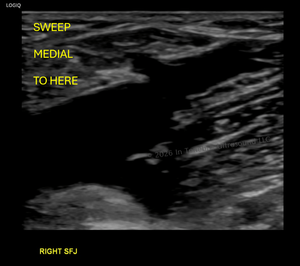
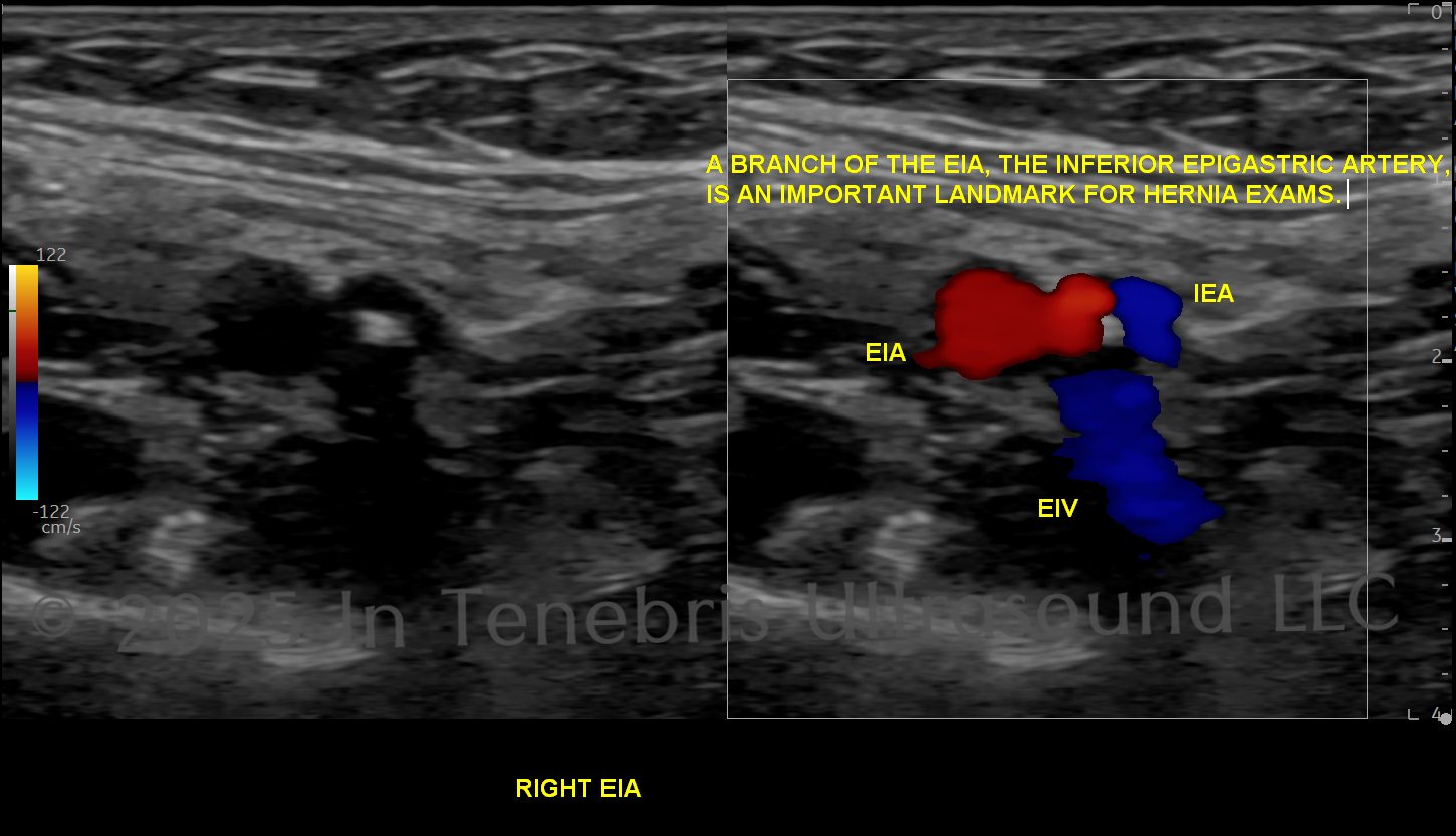
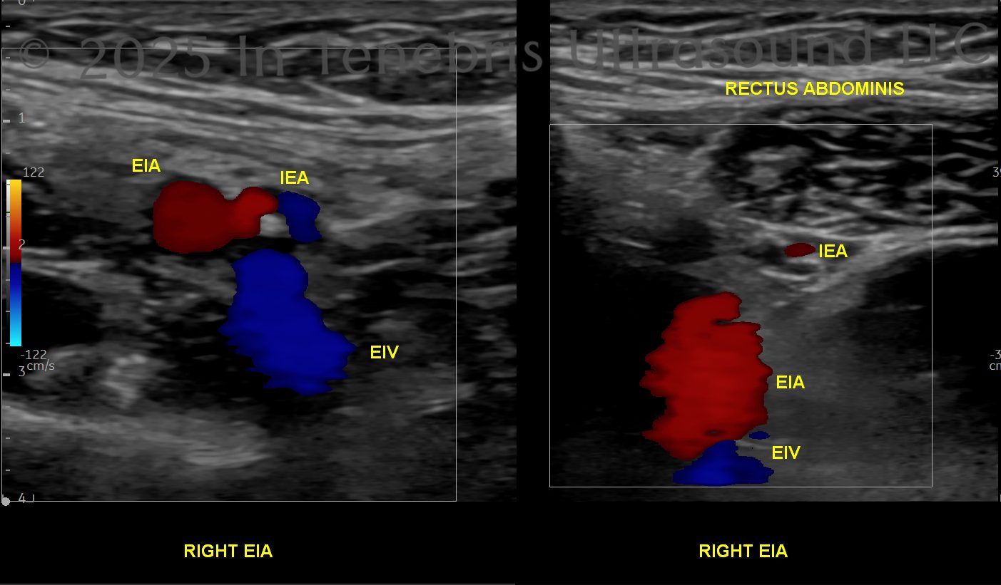
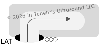
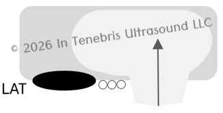
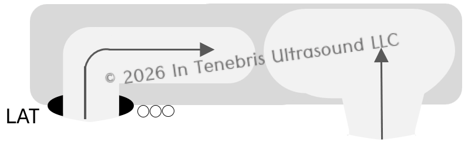
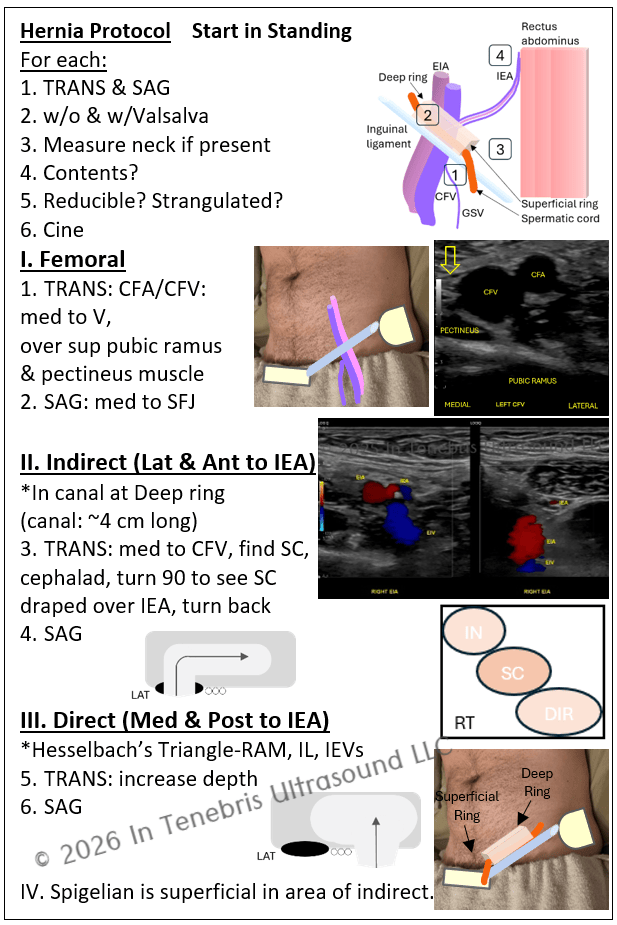
1. Femoral 2. Indirect 3. Direct 4. SpigelianLocation to assess for femoral herniaHere is where to look for both Indirect and Direct hernias: Indirect will be Lat to IEA, Direct will be Med.The IEA travels cephalad and superficial to join the rectus abdominis.Indirect hernia (looks like an “I”) — For direct and indirect hernias, this is the golden image: start here. The hernias will present on either side of the Inferior Epigastric Artery.Direct hernia (looks like a “D”) — For direct and indirect hernias, this is the golden image: start here. The hernias will present on either side of the Inferior Epigastric Artery.When both hernias are present, this combination is called “Pantaloons Hernia” and that’s one of my favorite things!