
Please See Core Concepts First!
They are, in fact, Physics.
Resolutions: The Foundation of Physics
Everything in sonography physics comes back to resolutions. Resolutions determines how well we can see. Resolutions are the foundation of image clarity and diagnostic accuracy.
4D
Ultrasound is a unique modality with a special ability: we can image in 4D. What does “4D” mean? That means we scan in four dimensions. We scan 3-dimensional structures. What are the three dimensions?
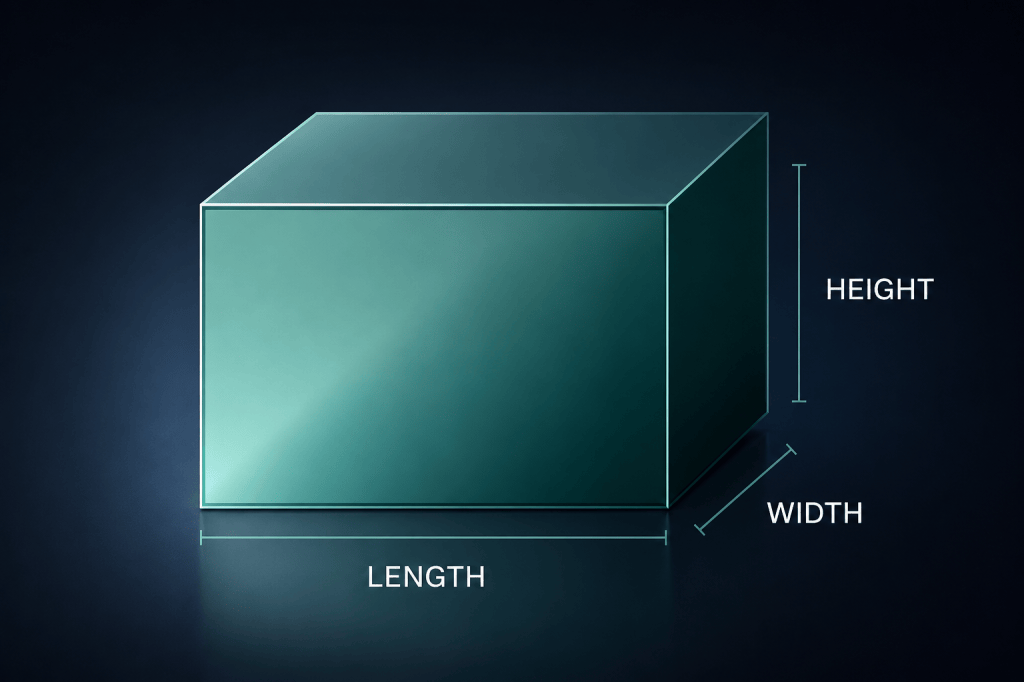
We always scan in two planes to see through all three dimensions.
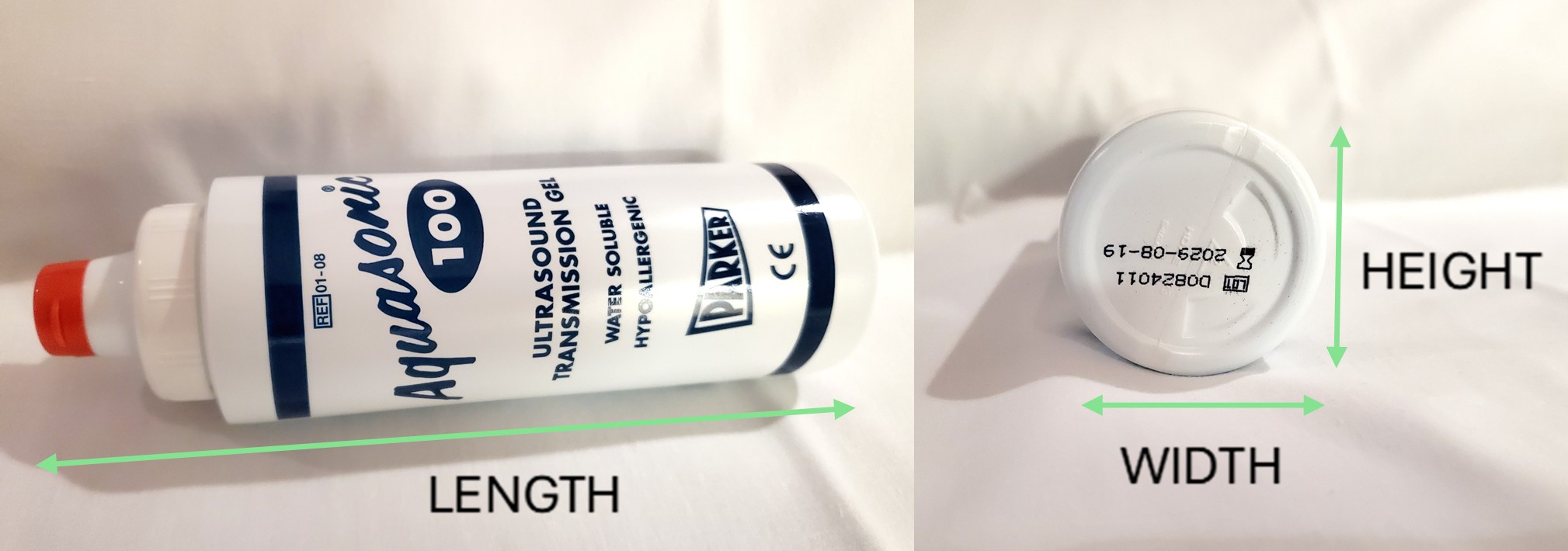
What, then, is the fourth dimension?
Time!
- We scan in two planes: a longitudinal plane (sagittal or coronal) and transverse.
- We scan in real-time and frame rate is important so we can accurately see how the structures lie in relation to others.
- If our temporal resolution is too poor, the machine will lag, stutter, and lose sensitivity to color and spectral Doppler.
Transducer → Beam-Shape → Spatial Resolutions
The shape of the transducer creates the shape of the sound beam.
The shape of the sound beam determines the resolution of the images.
The resolution of the images determines the clarity of the picture, the diagnostic accuracy, the removal of various artifacts, and helps distinguish borders.
Resolution is everything.
The smaller the beam is in its three dimensions, the better the sound beam can fit between the structures we are wanting to resolve.
The better the sound beam can fit between the structures we are wanting to resolve, the better we can resolve them and see them as distinct entities.
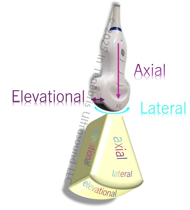
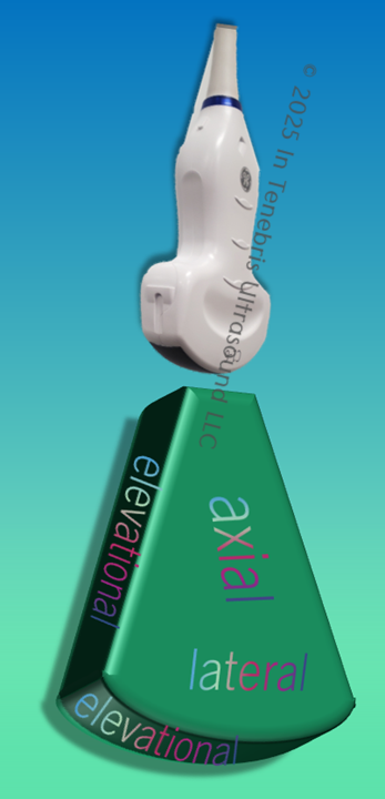
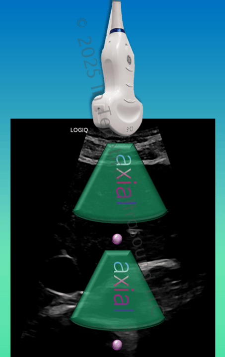
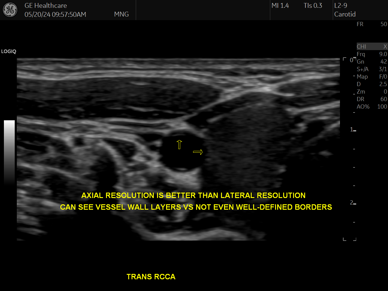
The height of the transducer (technically the thickness of the crystals in the transducer), in addition to the pulser, creates the spatial pulse length which creates the axial resolution. Remembering that the axial resolution originates from within the probe and projects out from the probe is helpful in your studies.
The length of the transducer face creates the beam width which determines the lateral resolution. I like to think of the lateral resolution (lateral means “side” and another name for this resolution is side-to-side) as the resolution that extends from one’s side to the other like a waist. Lateral resolution is best at the focal zone, which I like to think of as a belt, narrowing the waist.
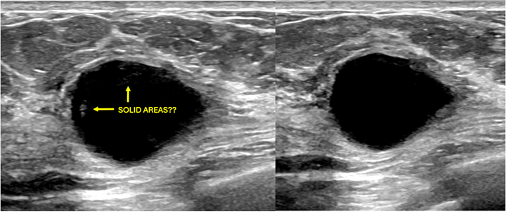
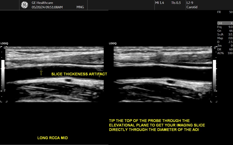
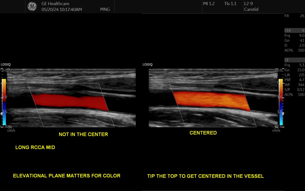
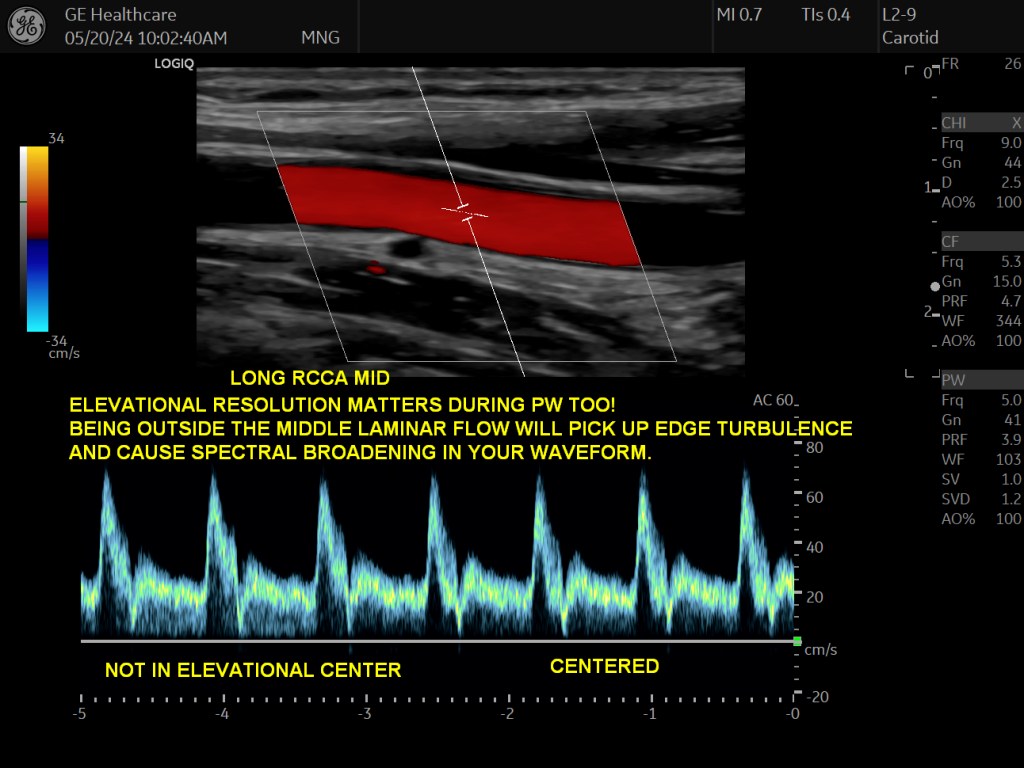
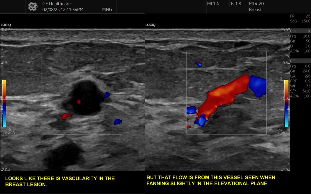
The width of the transducer face creates the beam thickness which determines the elevational resolution. We want the elevational resolution to be paper-thin. Just like an image is printed on a piece of paper, if the elevational resolution is poor, we can see text on the other side of the page which makes seeing our text or image difficult.
The 5 Resolutions
3 Spatial:
- Axial
- Lateral
- Elevational
Others:
- Temporal
- Contrast
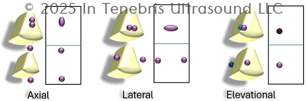
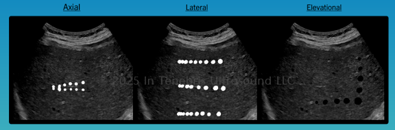
An ultrasound system must be able to resolve two pixels in order to be able to show them as separate and distinct, or else the two dots will appear as one dot.
Resolution simply means to show as two separate things.
The sound beam, in essence, must be able to fit between the two dots to resolve them.
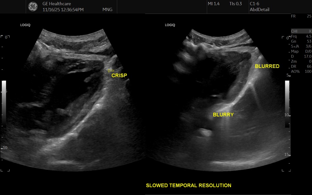
And then, the machine’s processing speed must be able to keep up in order to resolve the frames as you scan in real time.
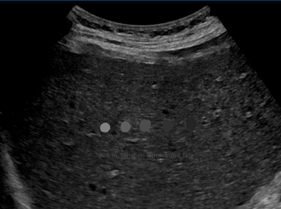
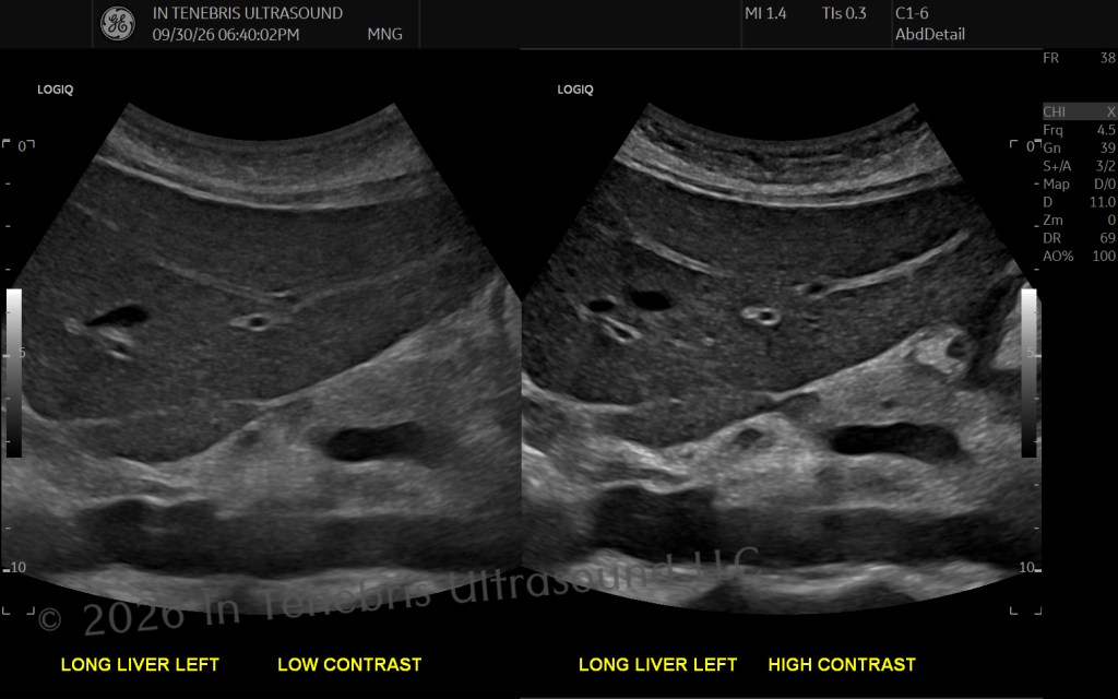
Finally, the shades of gray have to be different enough to show variances in the tissues being assessed.
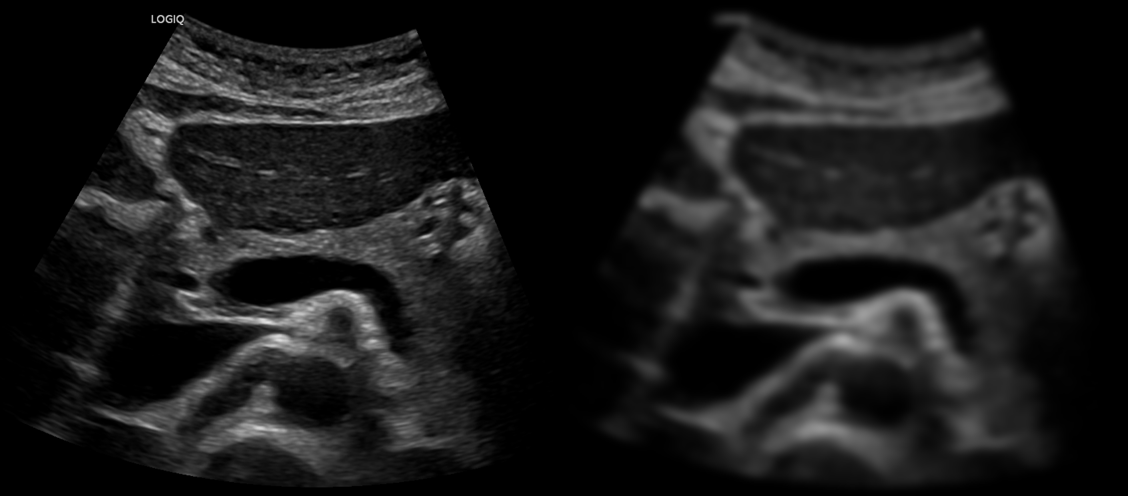
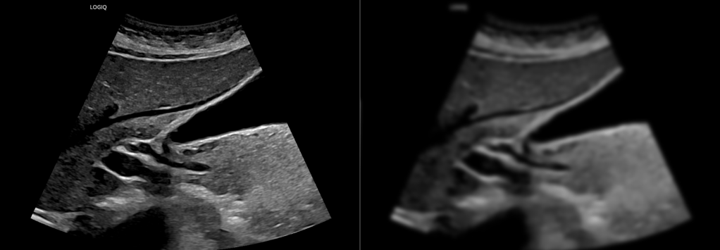
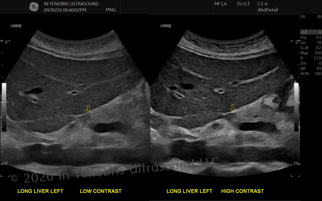
Take a look at the resolutions testing phantoms illustration. Seeing how the resolutions fail helps drive home the concept.
Axial Resolution
Superficial to Deep
Ultrasound that can resolve pixels to form images is pulsed wave (as opposed to continuous wave which only makes a spectral waveform line, almost like an electrocardiogram tracing). The shape of the beam is a single short pulse that travels from the transducer and away. The pulse must fit between two dots to resolve them.
Axial resolution is the parameter that we as sonographers can effect the most.
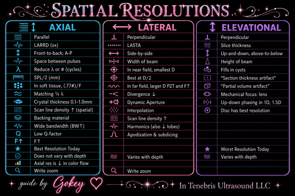
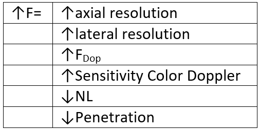
Axial resolution is the spatial pulse length divided by two. The spatial pulse length is determined by the number of cycles and the wavelength of the sound beam. The wavelength is inversely proportional to the frequency. As nearly everything in ultrasound physics comes back to resolution, the majority comes back to frequency. This is why. We effect frequency effects wavelength effects spatial pulse length effects axial resolution effects image quality. More notes and relationships on axial resolution follow.
Lateral Resolution
Side-to-Side
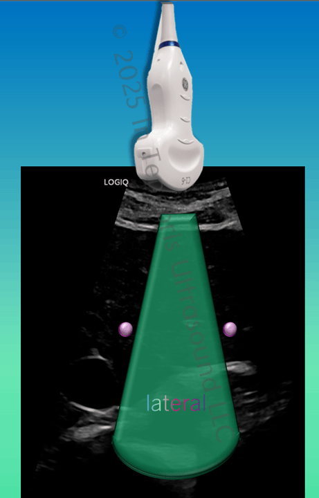
Lateral resolution corresponds to the waist of the ultrasound beam—the thinner the waist, the better the resolution. We mostly adjust the lateral resolution by adjusting the focal zone location on machines that do not run all foci all the time. More notes about lateral resolution follow.
Elevational Resolution
Toward Me; Away from Me
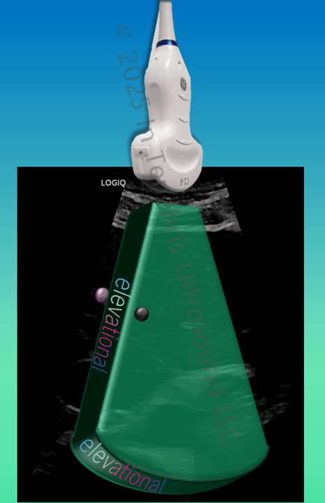
This is the resolution that is not in the 2-dimensional image. This is the dimension that, if you were to put on 3D glasses, would appear to jump out at you. Elevational resolution corresponds to the slice thickness of the ultrasound beam: think of the thickness of the crust of a slice of pizza. Our image should be one sheet of paper, but sometimes we can see the ink on the next page through our current page (like seeing a drawing that is on a page behind the sheet of paper, only we do not want to see that drawing)! Elevational resolution is especially noticeable when working with fluid-filled areas and seeing tissue—is the tissue within or behind the structure? Additional examples will follow.
Temporal Resolution
Frame Rate
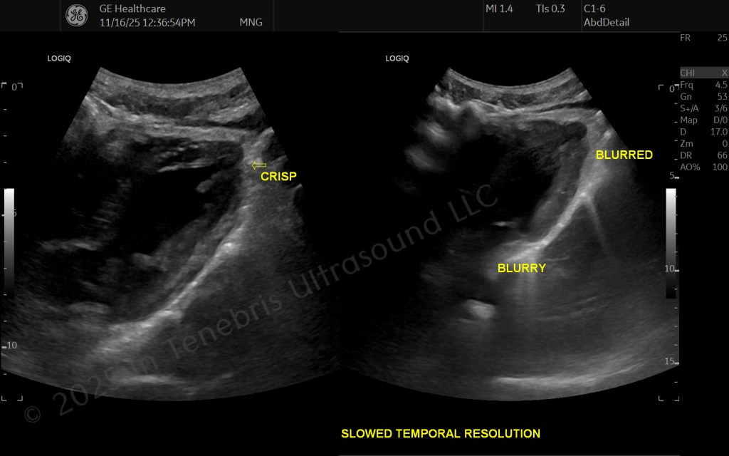
Temporal resolution is not just helpful to see a fetus moving around mom’s womb for a bonding moment. Temporal resolution helps us detect accurate color flow and blood flow velocities to identify pathology. Temporal resolution helps us safely guide interventional procedures in real-time. Temporal resolution helps us accurately measure heartrates. If temporal resolution is degraded enough, even fanning through the area of interest will blur the image on the screen. Recall that the Frame Rate is shown on the screen layout.
Contrast Resolution
Shades of Gray
Contrast resolution helps us see the different shades of gray between normal anatomy and abnormal anatomy. Pathological lesions can be a subtle shade different, and if they are not different enough (or even not at all), we will not be able to see them as separate from the surrounding normal tissue.
Application of the Resolutions to Measurements
The three spatial resolutions directly effect the accuracy of the measurements that we take. Your understanding of how which resolution influences which measurement will empower you to use your physics to optimize your borders to be able to visualize where to measure. Let us take a look.
The ovaries are highlighted in purple:
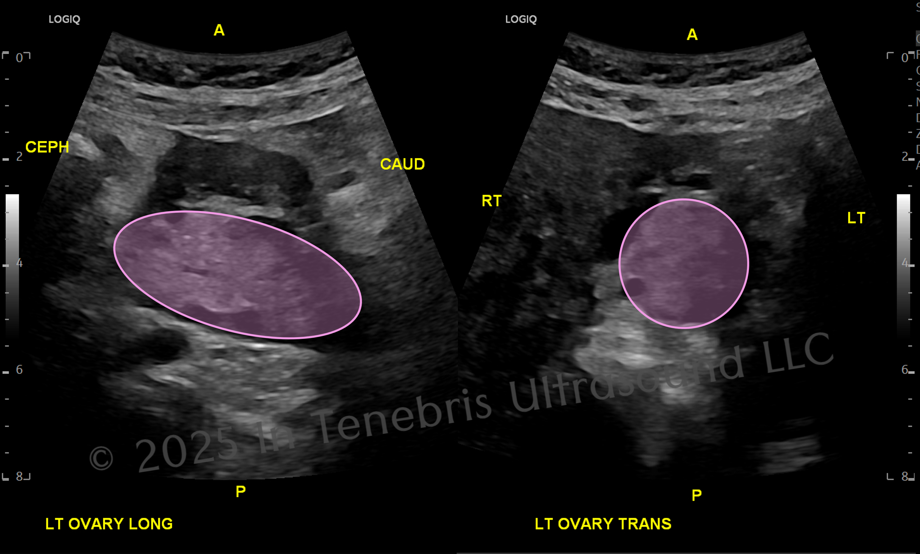
Orient yourself to these ovary images. Anterior (A) to posterior (P), cephalad (CEPH) to caudal (CAUD), and RT (right) to LT (left) directions are demonstrated.
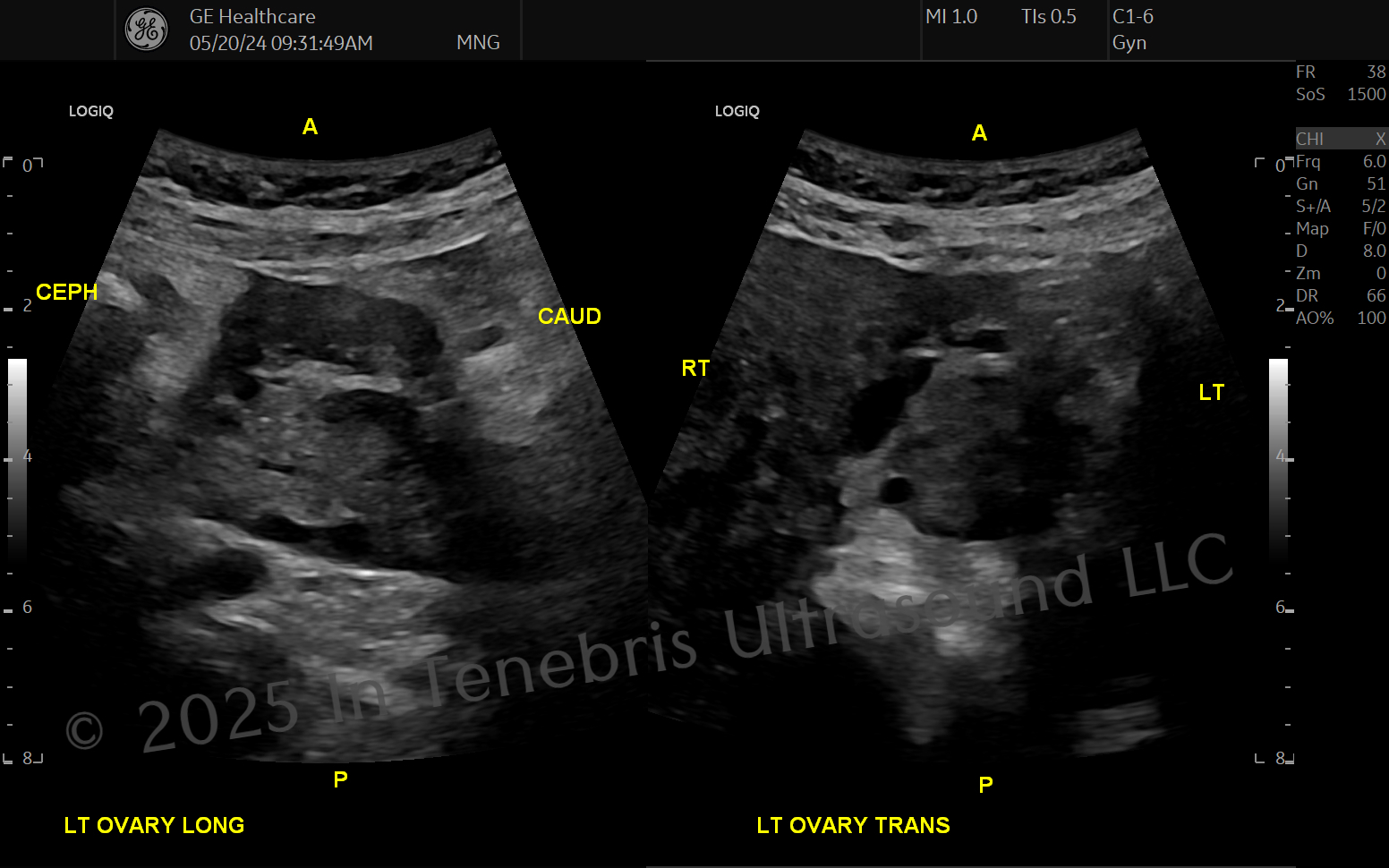
Note that both images are from A to P, meaning the ovary’s height is the same in both images. The height of the ovary does not change, and we are viewing the ovary from the same angle in both pictures.
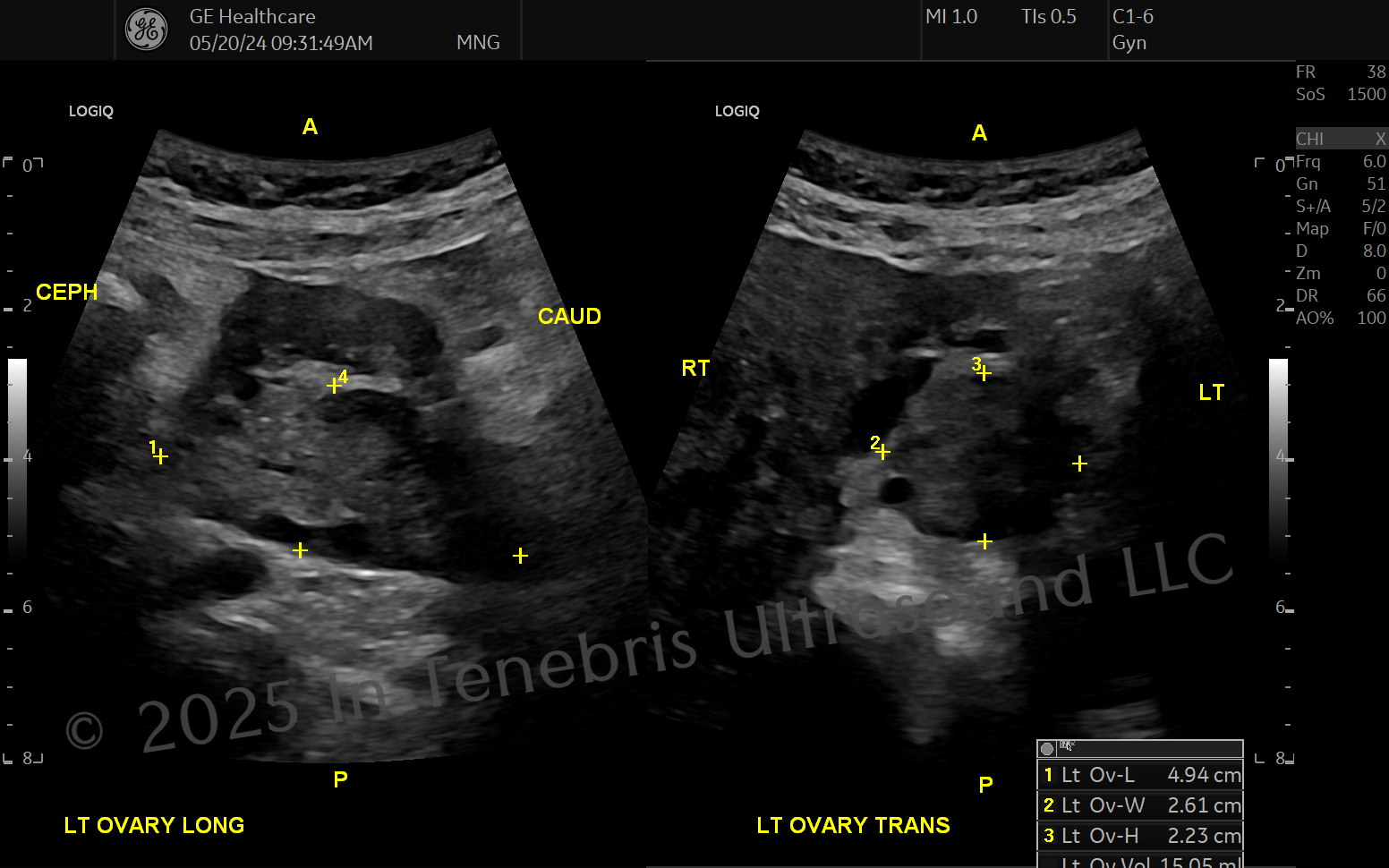
Consider these measurements and what you have now learned about axial, lateral, and elevational resolutions. Which resolutions effect length, width, and height?
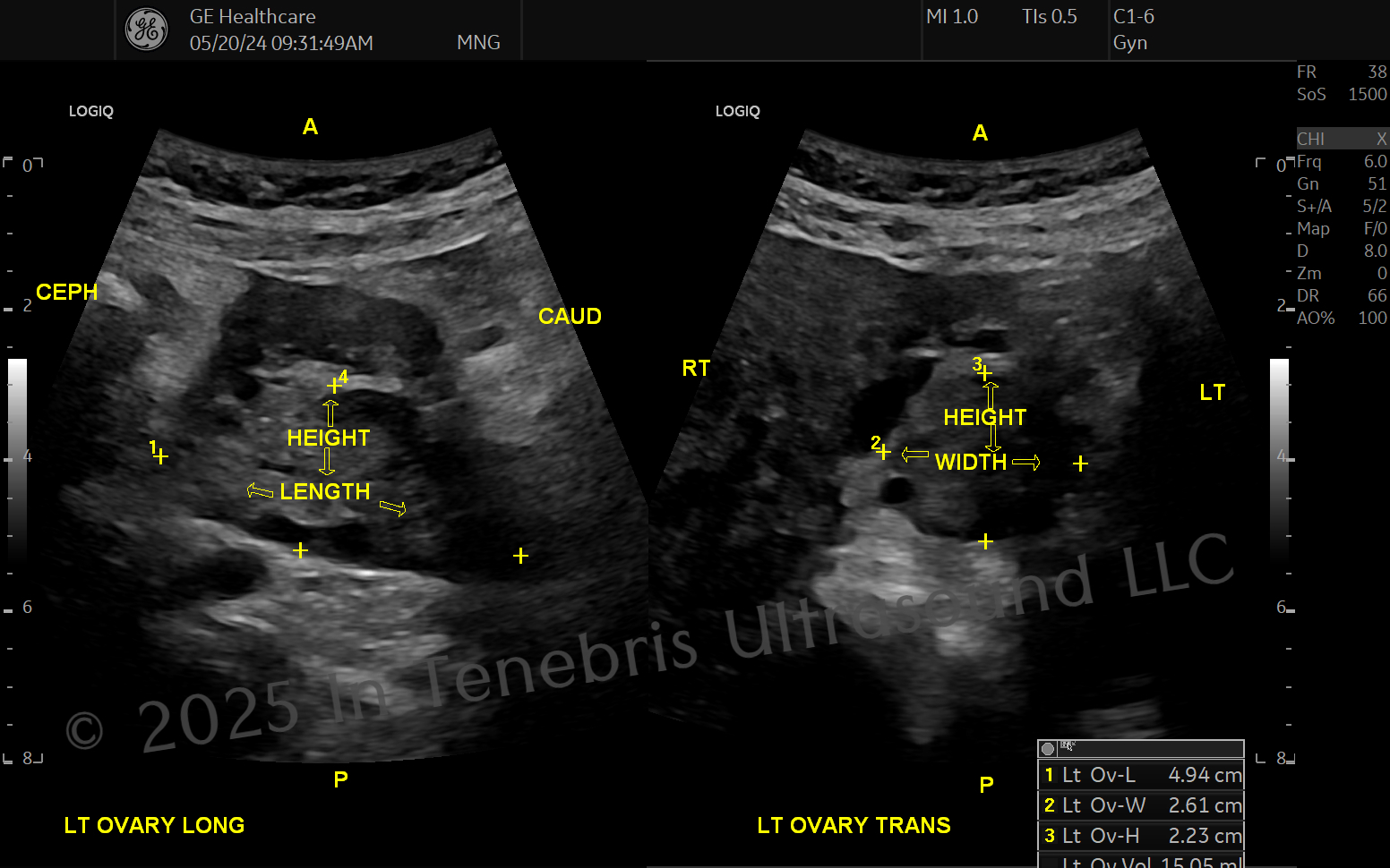
Axial effects height; lateral effects length and width! Where is elevational?
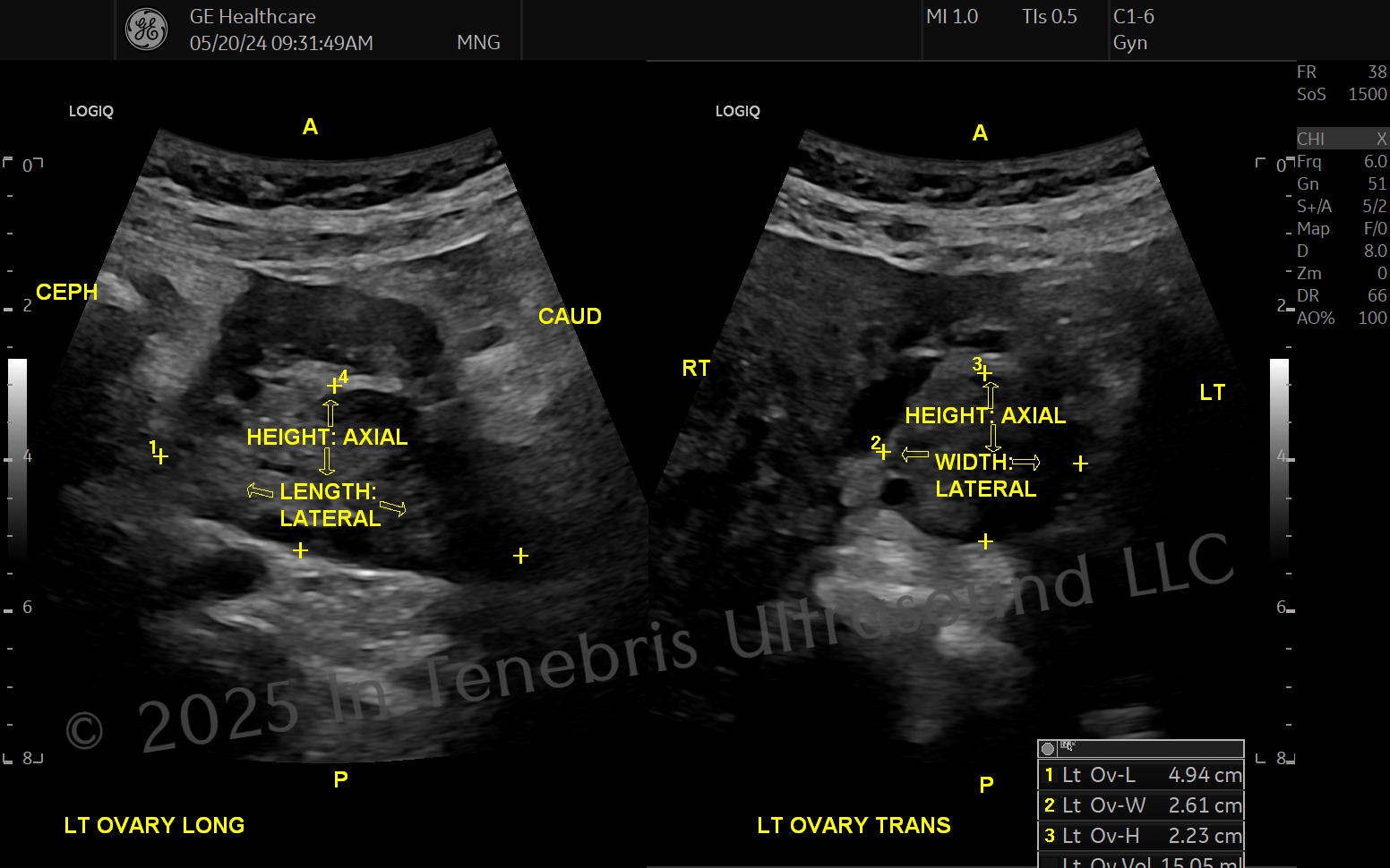
When optimizing borders for measurements, consider which resolution is affected. As you study your physics, pay attention to how you could make your axial borders pop, how you could make your lateral borders pop, and how you could make both pop! So where is elevational resolution? Think fluid. If there were a cyst in this ovary, and we needed to determine if the cyst is a simple cyst (benign, no follow-up required), has blood products (endometrioma, benign but problematic), or solid (worrisome, needs a workup), we would depend on our elevational resolution to determine if we saw any echoes within that space or not.
Physics of Resolutions
Sonography physics and instrumentation (SPI) almost always comes back to resolution and how each setting effects one of the resolutions. We optimize by improving resolution!
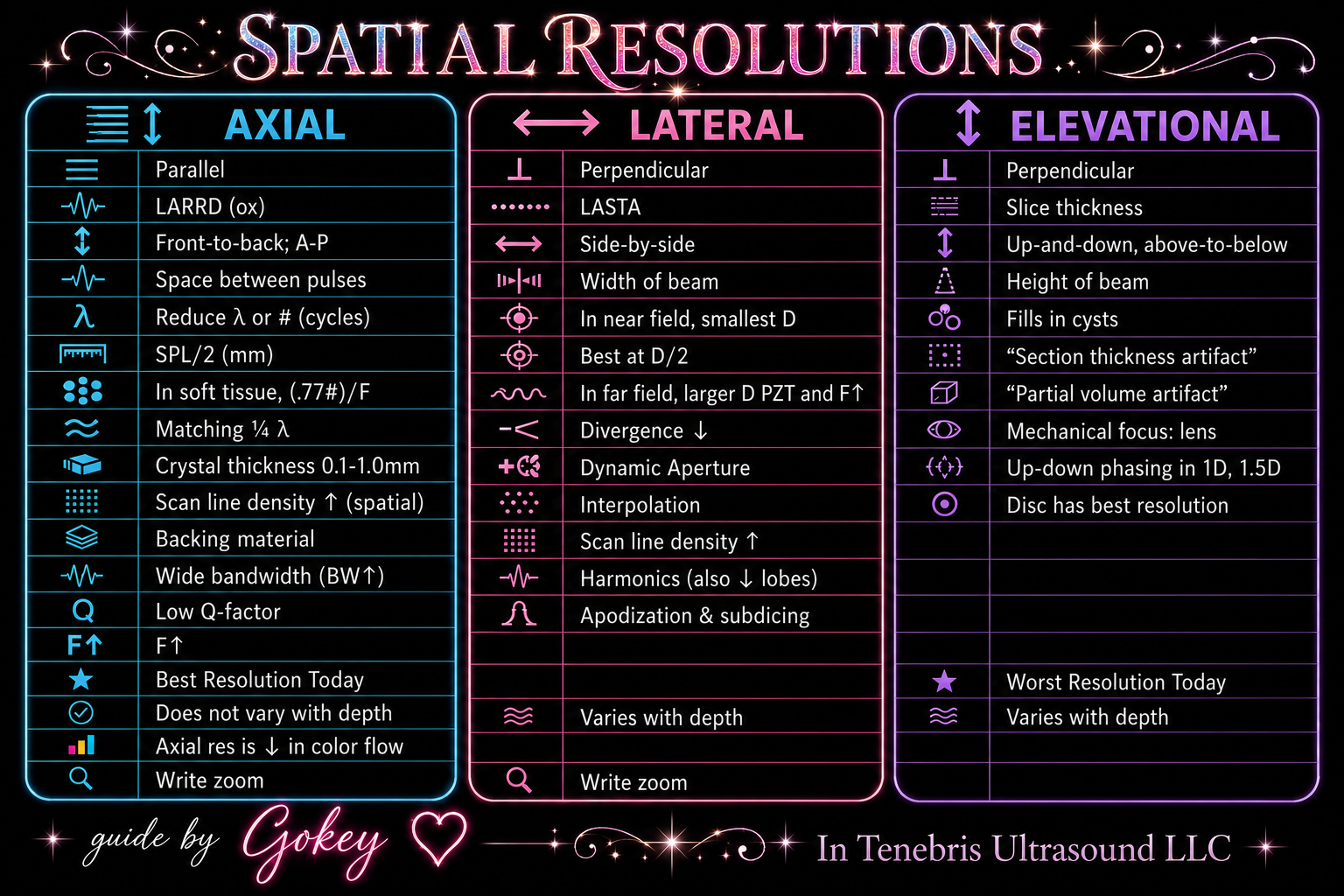
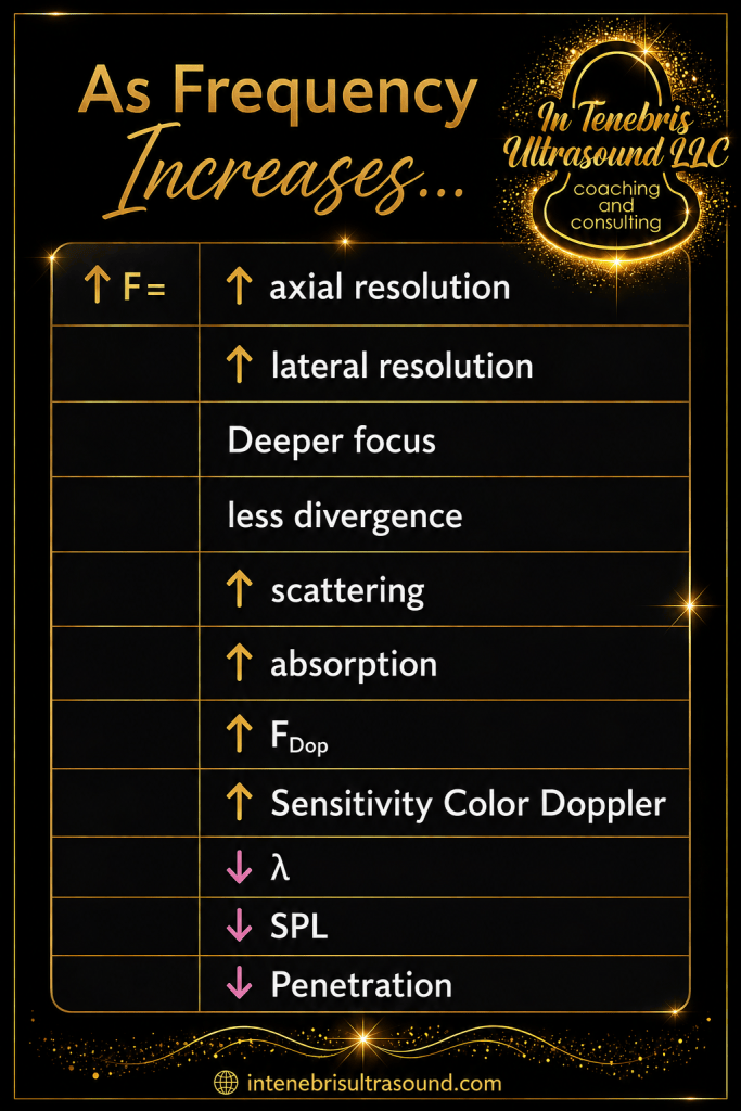
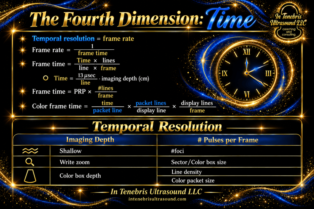
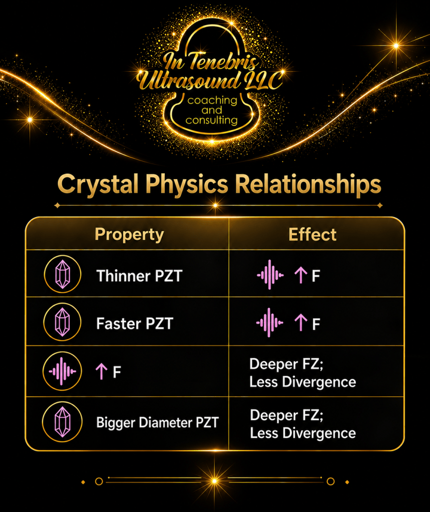
The following tables are summaries of notes from Miele (2013), Edelman (2012), and Davies (2015). Refer to these while you study physics. Pay attention to the “if, then” relationships. For example, if we increase frequency, then we reduce the wavelength. Keep going. If we reduce the wavelength, then we reduce the spatial pulse length (SPL). Almost there… If we reduce the SPL, then we increase the axial resolution! And there is the gold! These tables help illustrate these relationships for you and also highlight key concepts important for the SPI exam.
Two Tips to Learn Optimization Skills
- Check out your machine’s manual! The machine might even have optimization guides! You can often search the manual by clicking the F1 button on the machine.
- Play! See what happens when you push each button! Try the different settings under different types of scans.
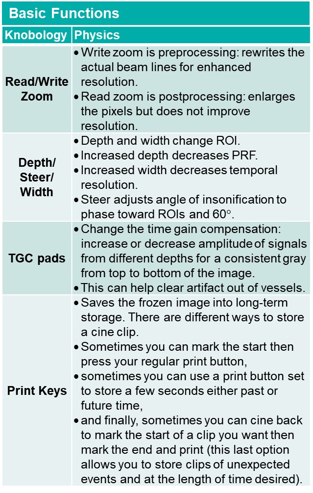
Different brands of machines will have different trademarked names for similar functions with different patented technology; for example, GE has SRI (speckle reduction imaging) whereas Philips has XRES (eXtended Real-time Enhancement System [GE Healthcare, 2012; Jago, Collet-Billon, Chenal, Jong, & Makram-Ebeid, 2002]). The effects remain similar unless a brand has a technology that others do not yet have.
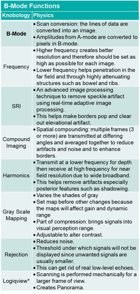
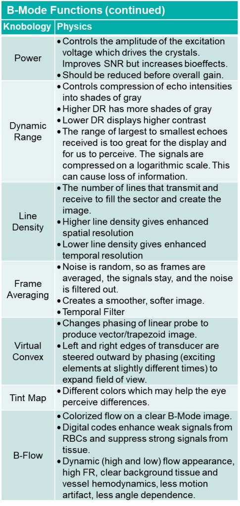
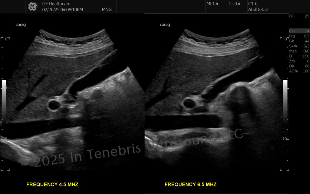
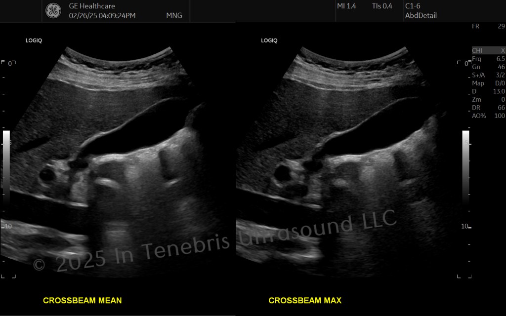
B-mode Optimization
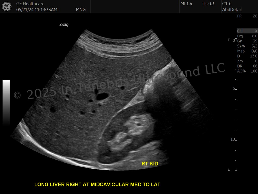
- Ensure a consistent shade of gray from near to far fields.
- Watch for shadows, reverberation, haze, and other artifacts that degrade the diagnostic quality of the image (hide too much tissue) or make areas look like pathology.
- Utilize your entire sector width by not cutting off one side of an area of interest while having empty space on the other side.
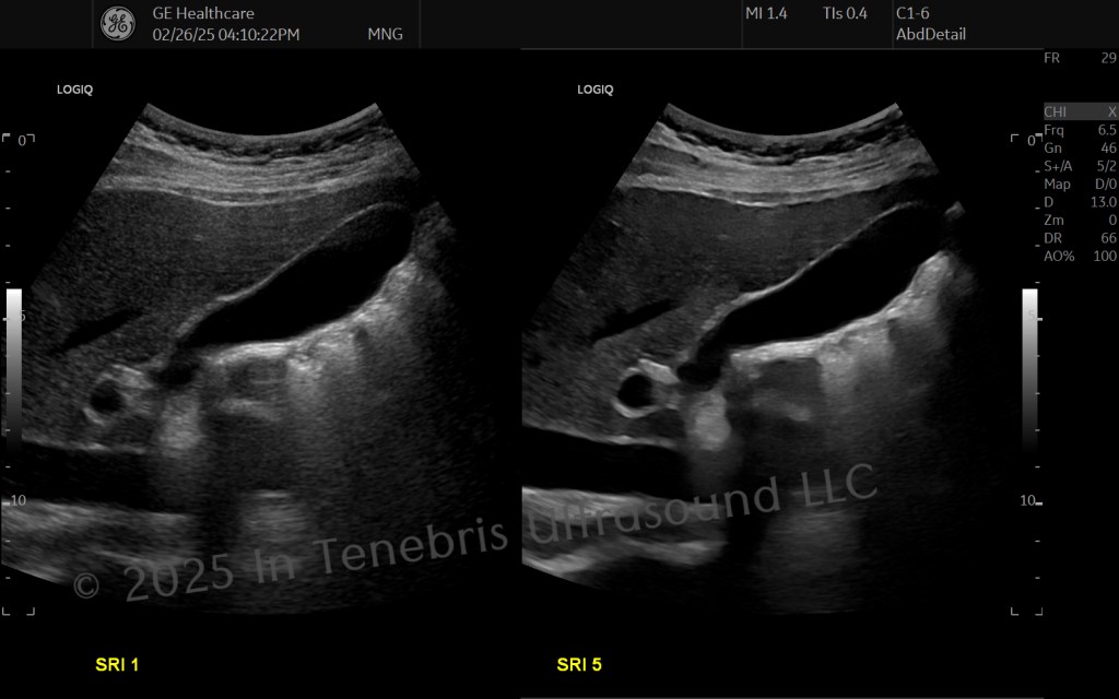
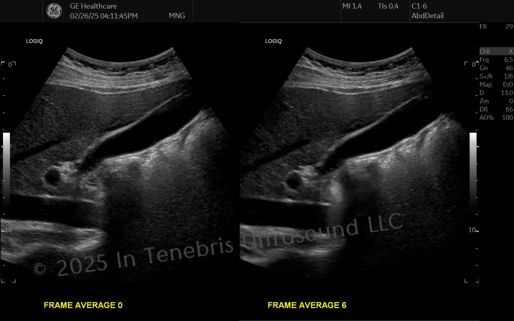
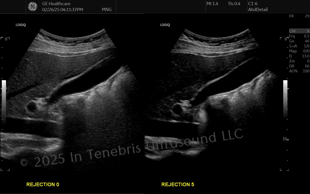
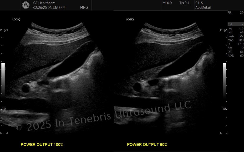
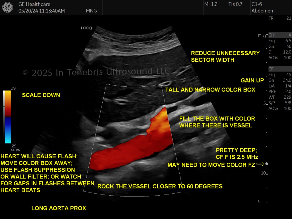
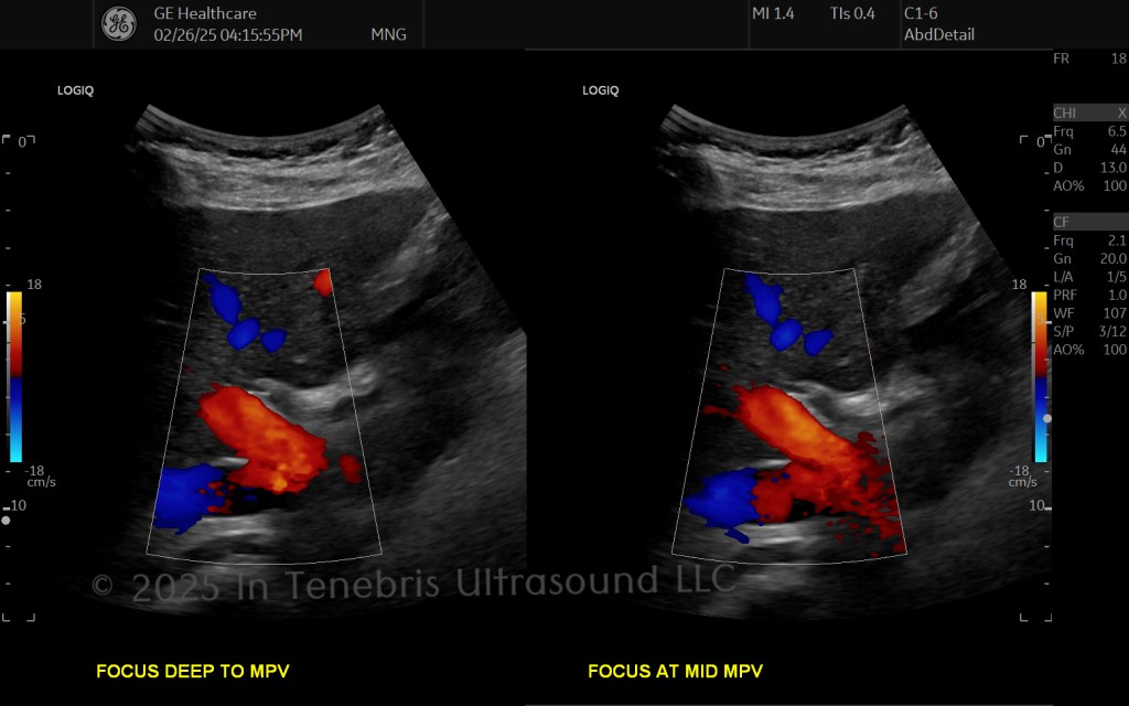
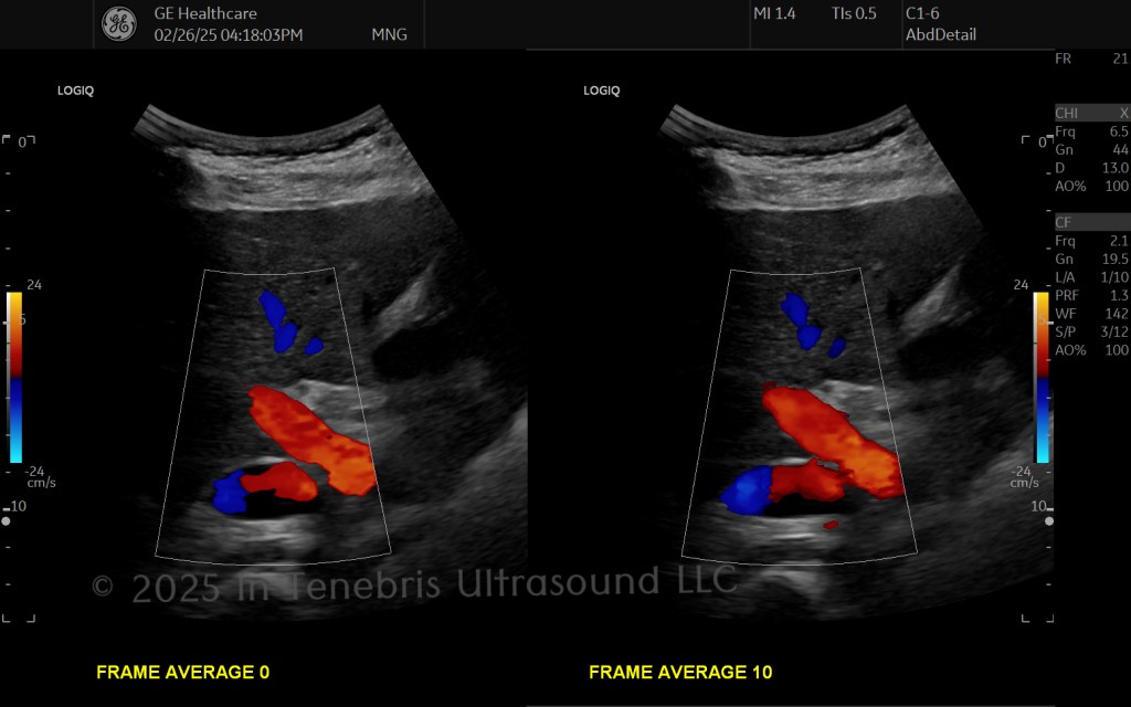
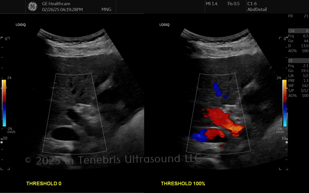
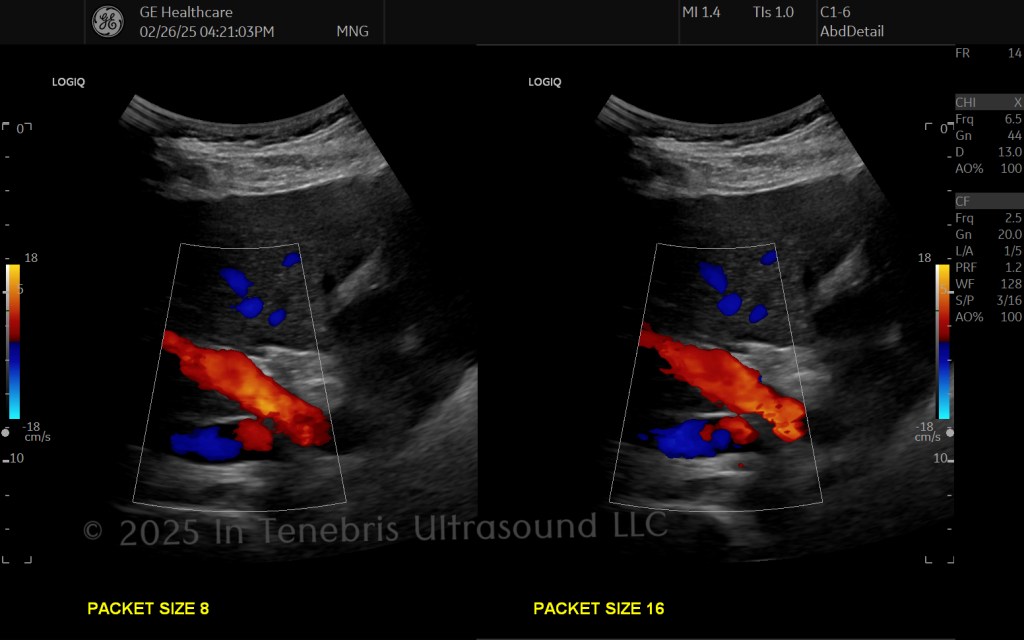
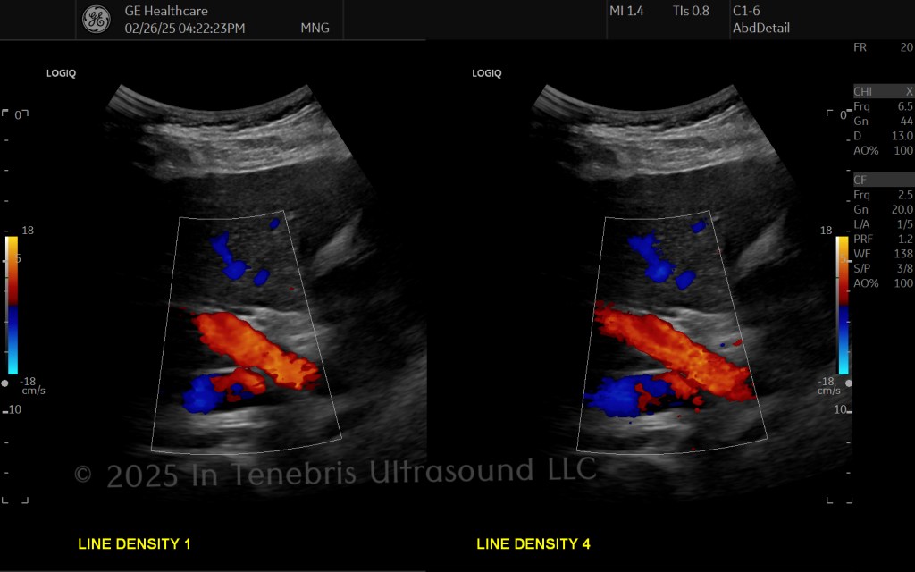
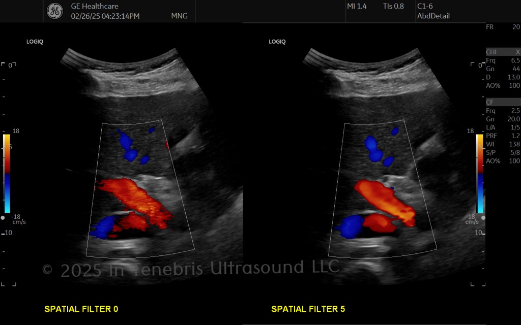
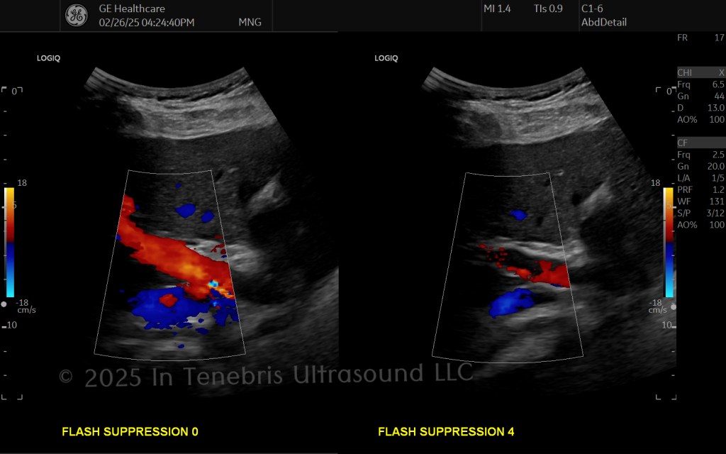
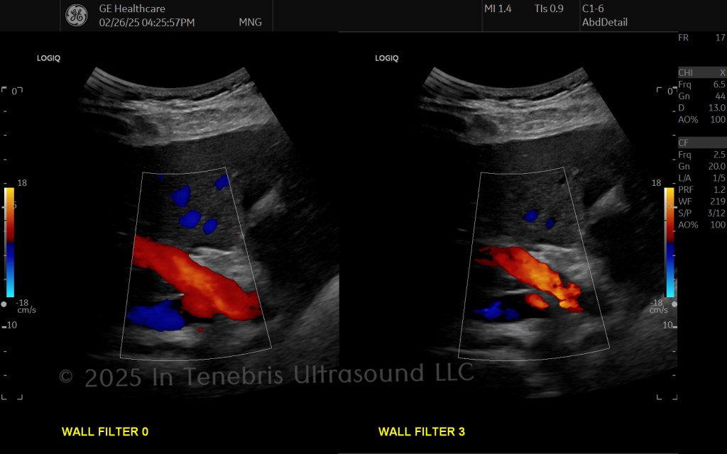
Color Flow Optimization
- Vessels should be as consistent shade of blue or red as possible.
- Fill the entire color box with color unless pathology interrupts the area.
- Do not blossom the color outside of the vessel.
- Do not post-process color flow gain.
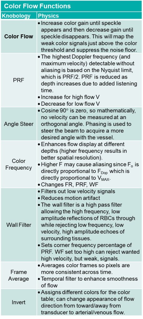
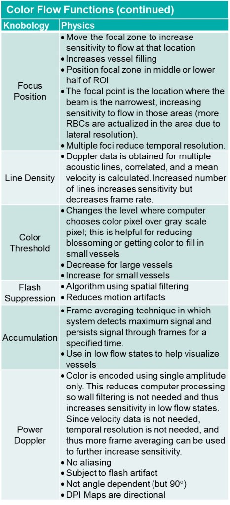
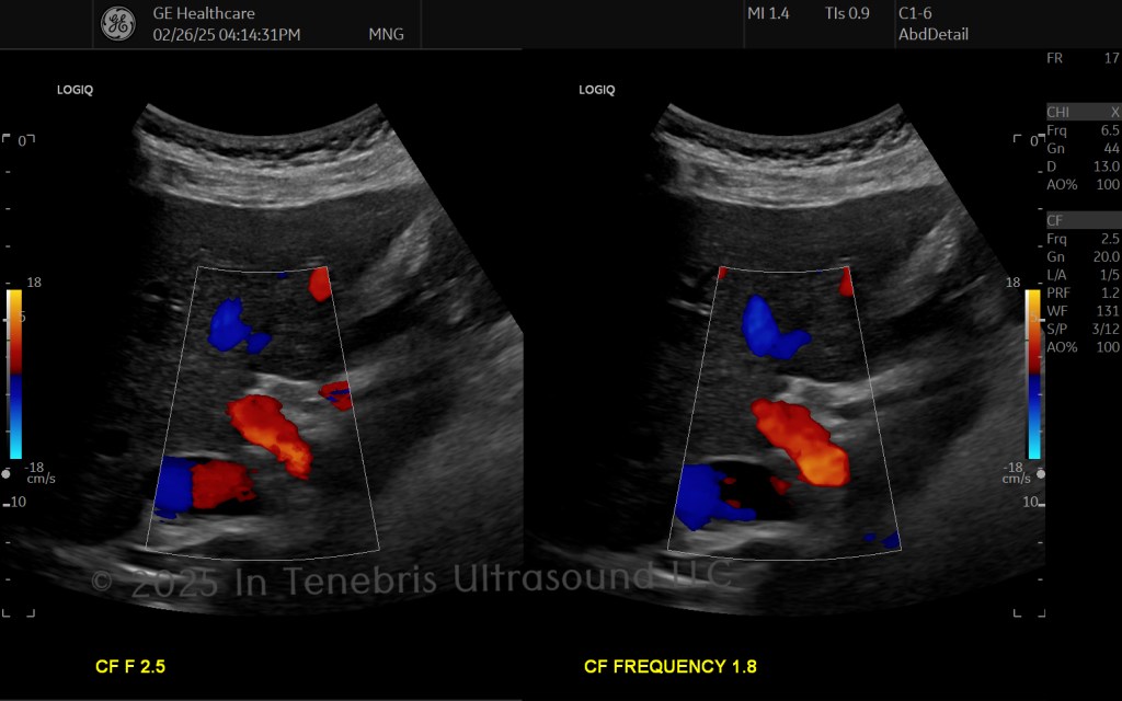
Pulse Wave Optimization
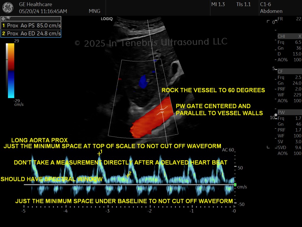
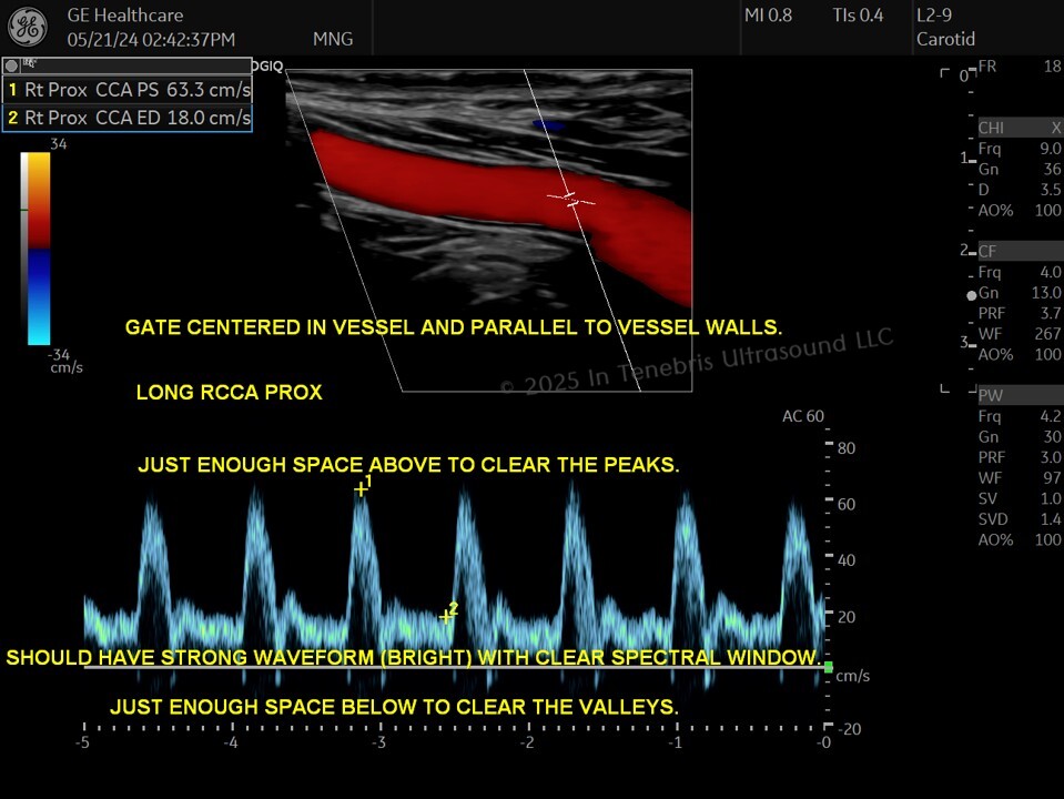
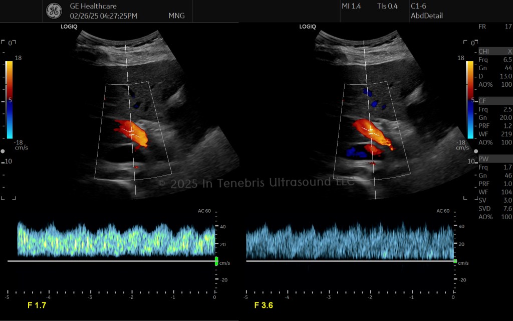
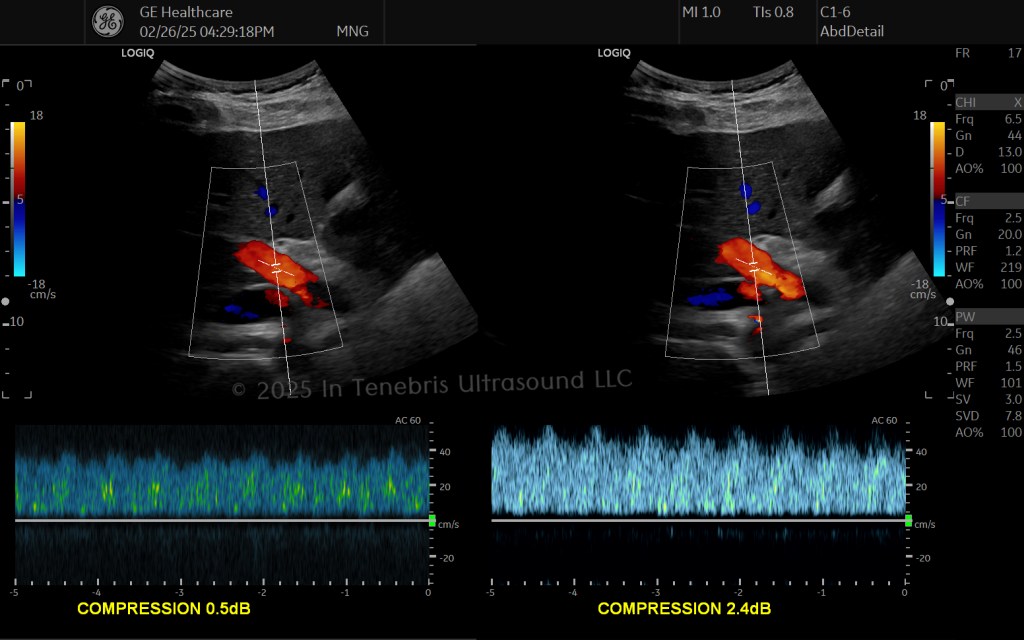
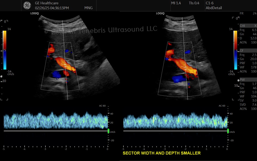
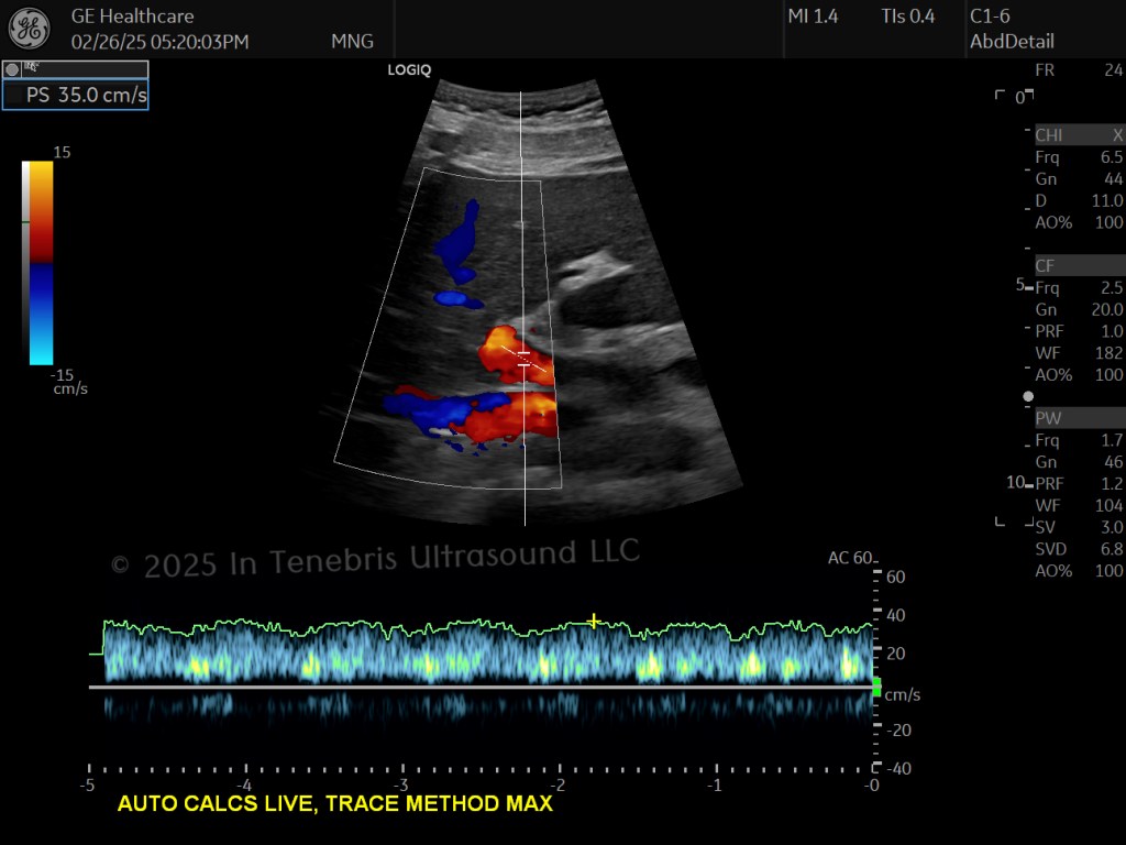
- Clearly show the top and bottom of the waveform by having scale (top of waveform) and baseline (bottom of waveform) set so that there is a small strip of black above the tallest peaks and a small strip of black below the lowest valleys. This is how we zoom in on the velocity measurements, ensuring the most accurate measurement possible.
- Set gain to show the waveform without showing unnecessary noise (echoes within spectral window and around the spectral line unless necessary for very low flow states).
- Do not post-process pulse wave gain. This will falsely change the peak velocity location.
- Spectral window should be clear unless the vessel truly has low flow during systole.
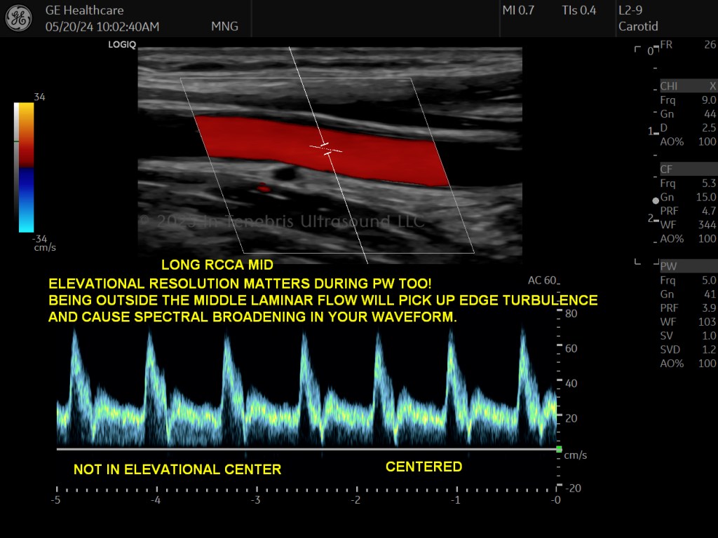
- Sample gate MUST be in the absolute center of the vessel (think all three planes, not just the two you can see in the image).
- Angle MUST be parallel to the vessel walls (or parallel to blood flow if flow direction is not parallel with the vessel walls). We are measuring the blood flow, not the vessel.
- Measure when at least three consecutive high quality cycles are obtained; do not measure directly after an arrythmia; try to not measure in an area of tortuosity (think velocity increases due to rollercoaster dynamics instead of stenosis).
- If serially evaluating flow velocities in a vessel, the recommendation is to maintain a similar Doppler angle between studies. This angle is usually set as 45-60 degrees (Oates et al., 2009; Tola & Yurdakul, 2006).
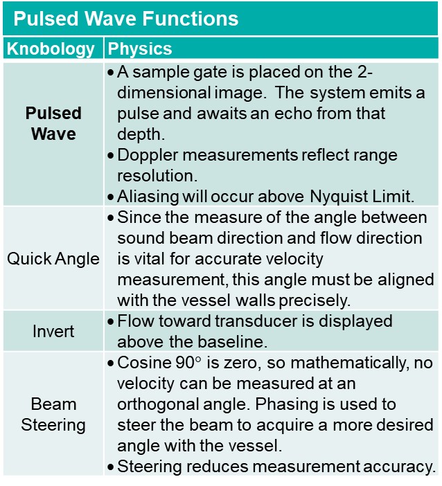
- Beam steering lowers velocity measurement accuracy. Rocking to obtain the required angle is ideal when feasible (Campbell et al., 2020; Oates et al., 2009; Russ et al., 2023; Steinman et al., 2005).
- Know where to measure (see the following definitions and explanations).
Pulse Wave Optimization: Knowing the Anatomy of the Waveform
Knowing the anatomy of a spectral analysis waveform is important for four primary reasons:
- Know if settings and technique are accurate
- Know where to measure
- Know what you are scanning (ECA vs. ICA, IVC vs. Aorta, etc.)
- Know how to describe your findings
Arterial Anatomy
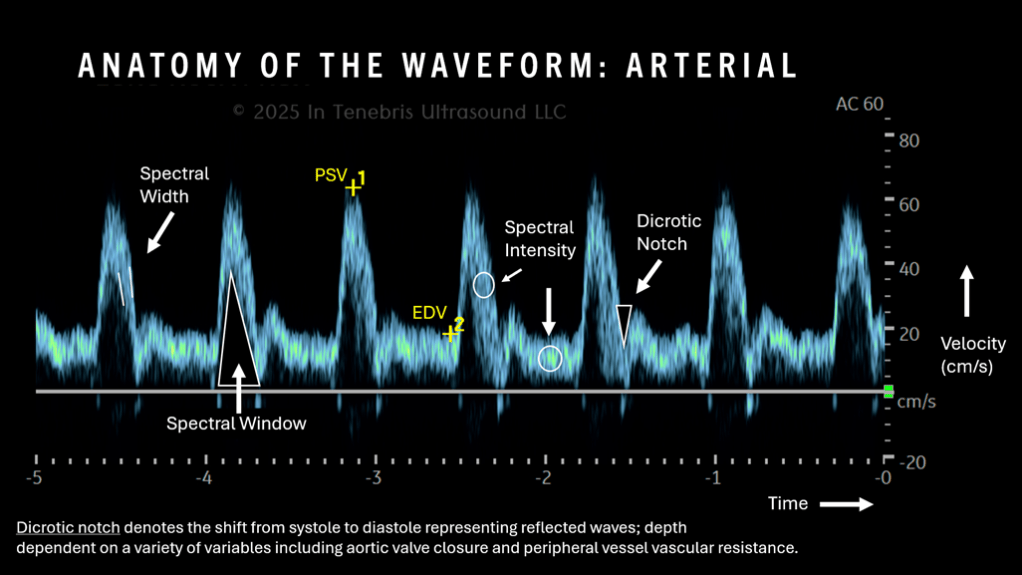
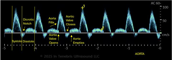
This is the physiology that causes the waveforms in the arteries that we record. Our lowest valley, end diastole, which we measure, is just before the systolic upstroke. End diastole corresponds to the lowest pressure in the arteries which happens when the aortic valve opens. Then the pressure increases in the arteries as blood flows into the aorta. We see the dicrotic notch, the dip in pressure when the aortic valve closes. Finally, pressure in the arteries falls as blood flows out of the aorta. All of the arteries we assess branch from the aorta; therefore, all of their waveforms are dependent on the pressure in the aorta.
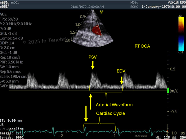
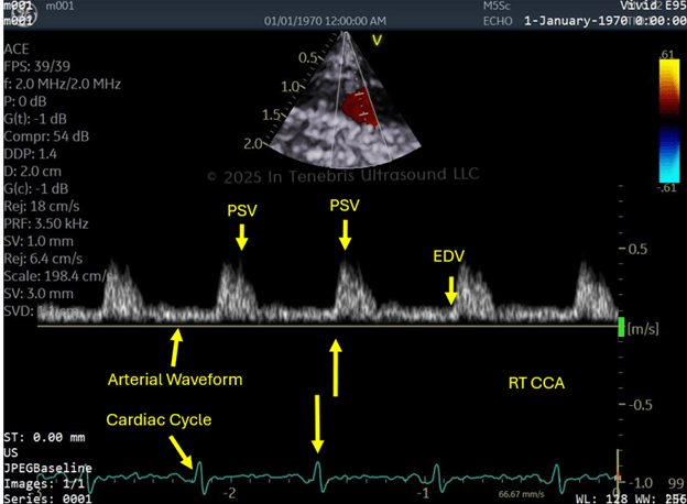
This image depicts the cardiac cycle from an electrocardiogram (ECG) tracing in a common carotid artery waveform in relation to the timing of the cardiac cycle in the heart (seen in green below the spectral waveform). From one peak to just before the rise of the next peak is a single cardiac cycle in each line. The heart pumps the blood out into the aorta, then we see the fill into the artery.
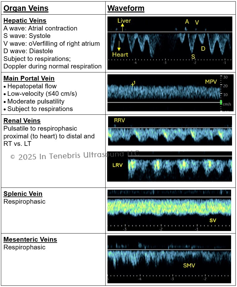
Venous Anatomy
Organs
Peripheral
- Spontaneous (calf vessels might be less so)
- Phasic
- Lower extremity venous (LEV) increase with expiration
- Upper extremity venous (UEV) increase with inspiration
- Vary in pulsatility
- UEV more pulsatile
- LEV some pulsatility may be normal but often indicates pathology
- Not continuous
- Will augment with compression or release of Valsalva
- Do not show reflux with Valsalva
See the UEV and LEV Protocols for examples!
Know What You are Scanning
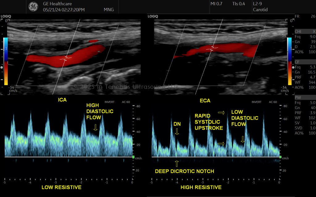
You can use your knowledge of arterial versus venous waveforms to help you determine which vessel you are interrogating. Additionally, knowing the differences in normal internal carotid artery (ICA) and external carotid artery (ECA) waveforms can help you distinguish which one you are in before you even take your first B-mode image (these are tricky at first). You can “drop a dirty Dop” and use PW without optimizing to get a quick sample of the waveform to determine what the vessel is without the intention of taking any spectral images yet. Use the Doppler as a tool, not just one more picture you have to take in your protocol. Remember too, color is also your friend! Try keeping CF on as you sweep from ICA to ECA and lengthen them out. The vessels are much easier to appreciate all lit up than darkened!
Know if Settings and Technique are Accurate
Now that you understand the anatomy of the waveform, you can appreciate if you need to adjust your technique and/or your settings. The first thing we should always ensure is that we are in the absolute center of the vessel that we are interrogating. This means not only in the center of the two planes that we can see (top-to-bottom and side-to-side), but also that pesky third plane that we cannot see! How can we tell if we are where we are supposed to be? We clear out our spectral window by tipping the top of the probe through the elevational plane (do this maneuver before adjusting gain: not only will you clear out the window, you will get a brighter, stronger spectral intensity. You will be more diagnostically accurate). Once your technique is accurate, ensure your settings are correct: baseline, scale, and gain must all be optimized properly so that your measurements are correct.
Know Where to Measure
There is a surprising amount of confusion about this amongst professional sonographers. You, however, will know the accurate way to measure because you understand the anatomy of the waveform and what your measurements mean.
Several studies define and describe peak systolic velocity (PSV) and end-diastolic velocity (EDV), including Chavhan et al. (2008), Kim et al. (2020), O’Shea et al. (n.d.-a), O’Shea et al. (n.d.-b), and Pellerito and Polak (2012). PSV is the tallest peak anywhere in systole. EDV is the velocity just prior to the next systolic upstroke.
PSV: The highest spot along the length region of the spectrum which correlates with the systolic phase of the cardiac cycle.
EDV: The EDV corresponds to the point marked at the end of the cardiac cycle (just prior to systole).
Peak Velocity (PV): The maximum positive Doppler shift during a cardiac cycle, used for portal veins (Gallix et al., 1997).
Know How to Describe Your Findings
The terms used to describe vascular ultrasound findings are standardized by the publication by Kim et al. (2020). This is a richly-illustrated article that I strongly recommend that you obtain for your reference.
- Arterial
- Flow Direction
- Phasicity
- Resistance
- Other terms (upstroke descriptors, sharp peak, spectral broadening, staccato, dampened, flow reversal)
- Venous
- Flow direction
- Flow pattern
- Spontaneity
- Other terms (augmentation, reflux, fistula flow)
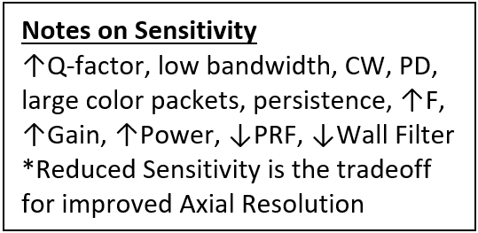
To Increase Sensitivity
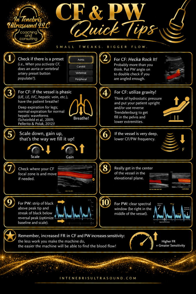
- Decrease scale (PRF).
- Turn up CF/PW gain, power.
- Reduce F for penetration; Increase F for FDop.
- Turn down B-mode gain (Threshold).
- Reduce threshold.
- Reduce wall filter.
- Use accumulation.
- Use power Doppler.
- Use CW.
To Improve Color Resolution
- Use correct box location for focus.
- Increase color packet size (Color packet size↑=↑SNR).
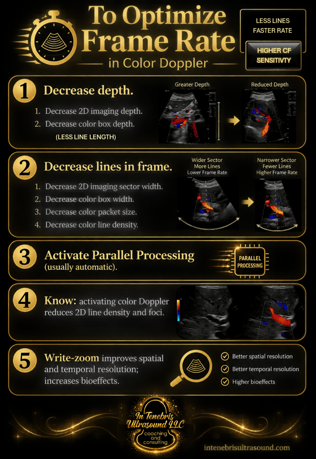
- Increase line density.
- Use frame average.
- Wall filter reduces flash artifact and slow flow.
- Flash suppression reduces flash artifact.
- Use spatial filter.
- What affects spatial resolution of color flow? Line density and Frequency.
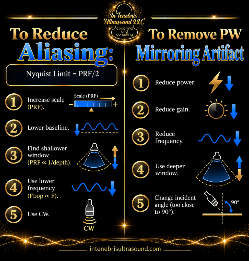
Artifacts
An artifact in ultrasound is something false in our image: a shadow, a brightness, an echo, etc., that does not reflect what is really in the body.
Sometimes artifacts are actually helpful.
A lot of our optimization work is to work around and/or eliminate artifacts to make sure that what we are seeing, and what we are showing our reading physician, is real.
Accidentally showing an area and making the area appear to have pathology is surprisingly easy.
Costly additional imaging may be ordered for a patient because a renal cyst was not properly cleared out during the survey images.
A patient may be sent to surgery because the CBD during a challenging emergency room (ER) scan was not cleared out.
Knowing your artifacts, knowing how to determine if what is being seen is indeed an artifact and not real, and knowing how to eliminate unwanted artifacts from your imaging is paramount.
Baad et al. (2017) provide a comprehensive and well-illustrated review of ultrasound artifacts that serves as a highly valuable reference for sonographers.
Combining Optimizations & Accurately Documenting Pathology
- Show in Survey Images
- Long
- Trans
- Measure
- Color
- Spectral
- When we identify pathology during the survey, we note to ourselves the two largest and most concerning representations of a single disease process. Then we document.
- Be sure when you do your survey of the organ you show the area of interest (AOI) in your sweep in both long and transverse planes. Show the area(s) in relation to major vessels whenever possible.
- Then show the AOI optimized: write zoom in*, frequency as high as possible*, speckle reduction*, edge enhancement, frame average, etc. (*Always do). Find the longest dimension for long, turn 90 degrees and document trans. Label long and trans relative to where they are in the sweep; i.e., Long Uterus LT and Trans Uterus Mid.
- Measure including any surrounding halo or capsule. Color can help identify borders.
- Scale down and gain up appropriately for color. Sweep through the AOI in two planes with color on to find relevant flow patterns. Remember that angles matter. Try PDI. Color patterns can help differentiate between concerning or likely benign growths (for example, liver focal nodular hyperplasia versus a liver adenoma). If you are scanning a superficial area, such as in the breast, subcutaneous tissue, or skin, lighten up pressure so that you do not compress small vessels.
- Doppler any significant vessels to determine low or high resistive patterns.
- For vascular studies, be sure to walk your Doppler gate through any areas of stenosis to find the highest velocity. Also document waveforms pre- and post- stenosis. Document plaque clearly.
- For vascular studies, when evaluating stents and graphs, be sure to document velocities pre-, proximal, mid, distal, and post- the intervention.
- Check out pancreas, liver, renal, female pelvis, scrotal, thyroid, and mesenteric Doppler for examples of pathology!
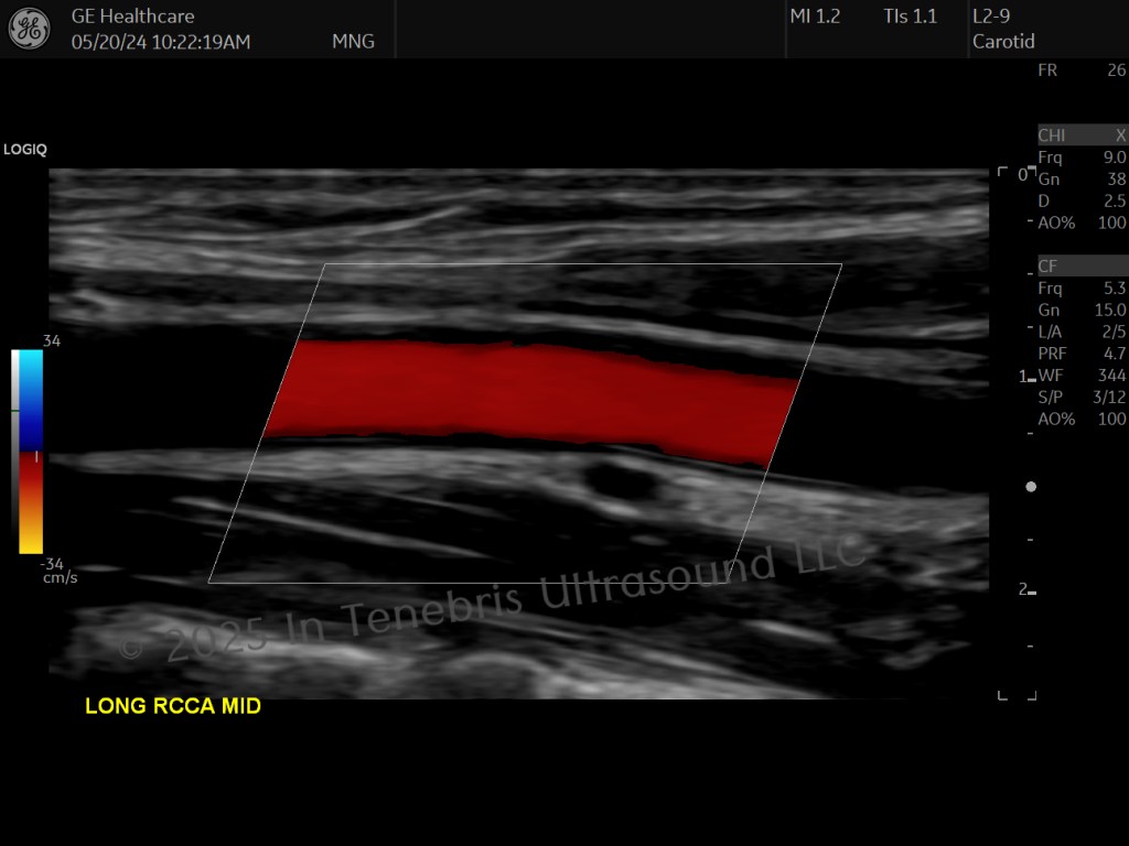
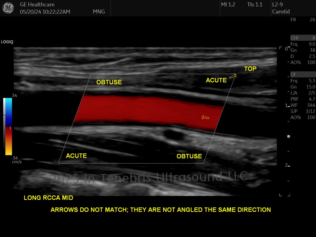
Guide to Color Box Steering with a Linear Probe
This concept is very confusing at first. If you are ever unsure, put pulse wave on and you will see the correct way the angle should be and the corresponding box steering. The box should be angled the same direction as the vessel.
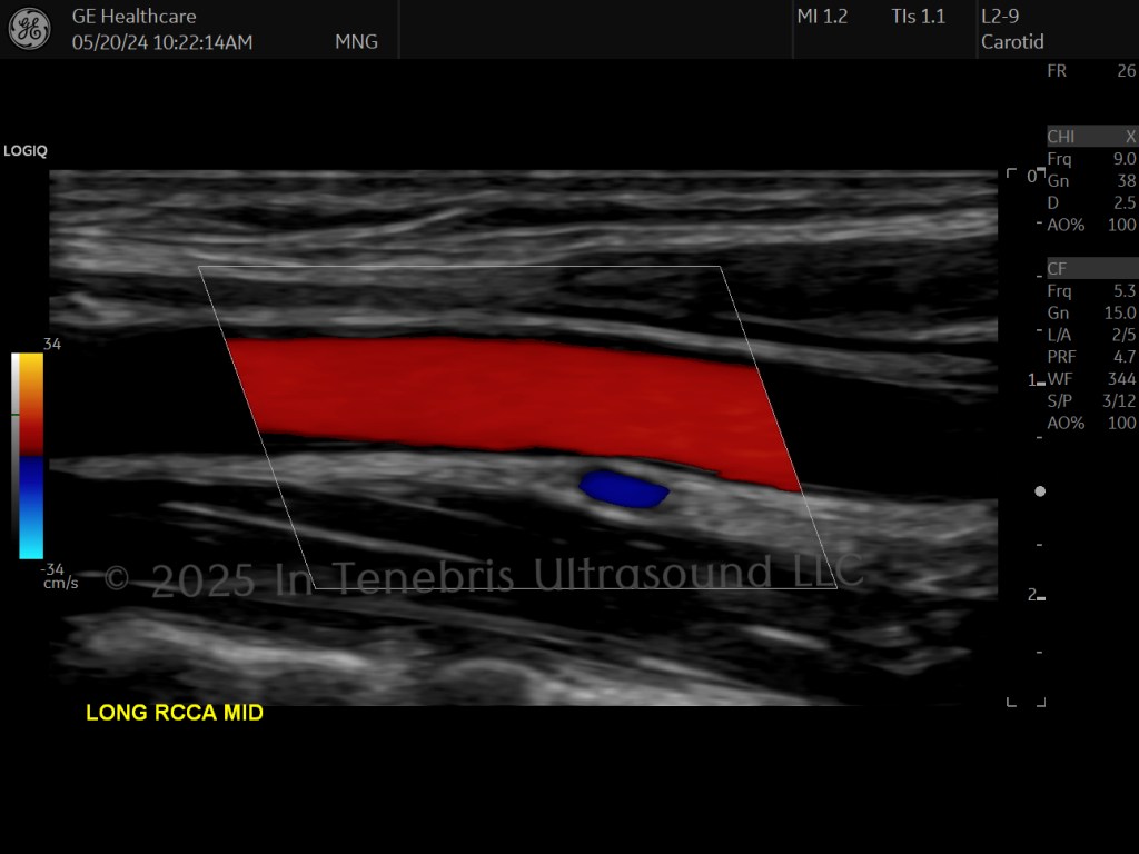
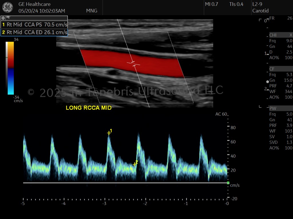
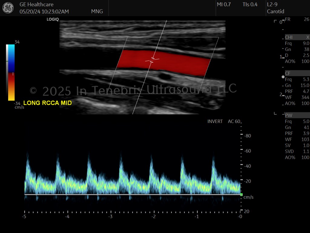
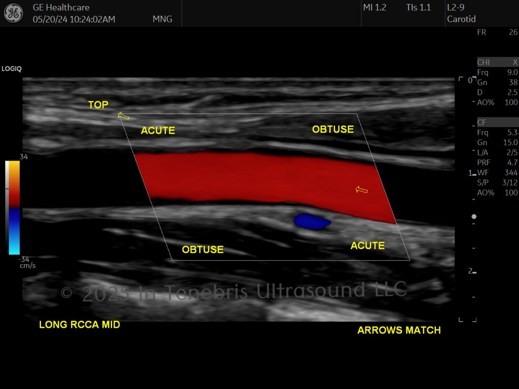
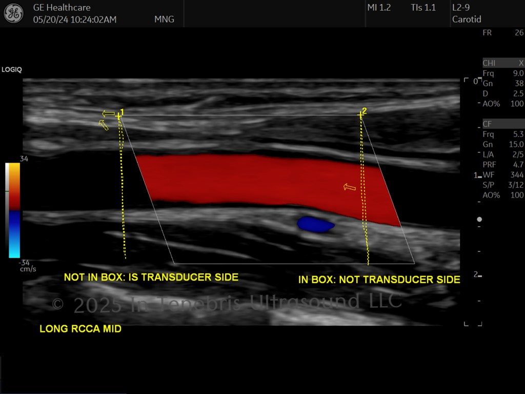
Guide to Determining Blood Flow Direction
Linear Probe
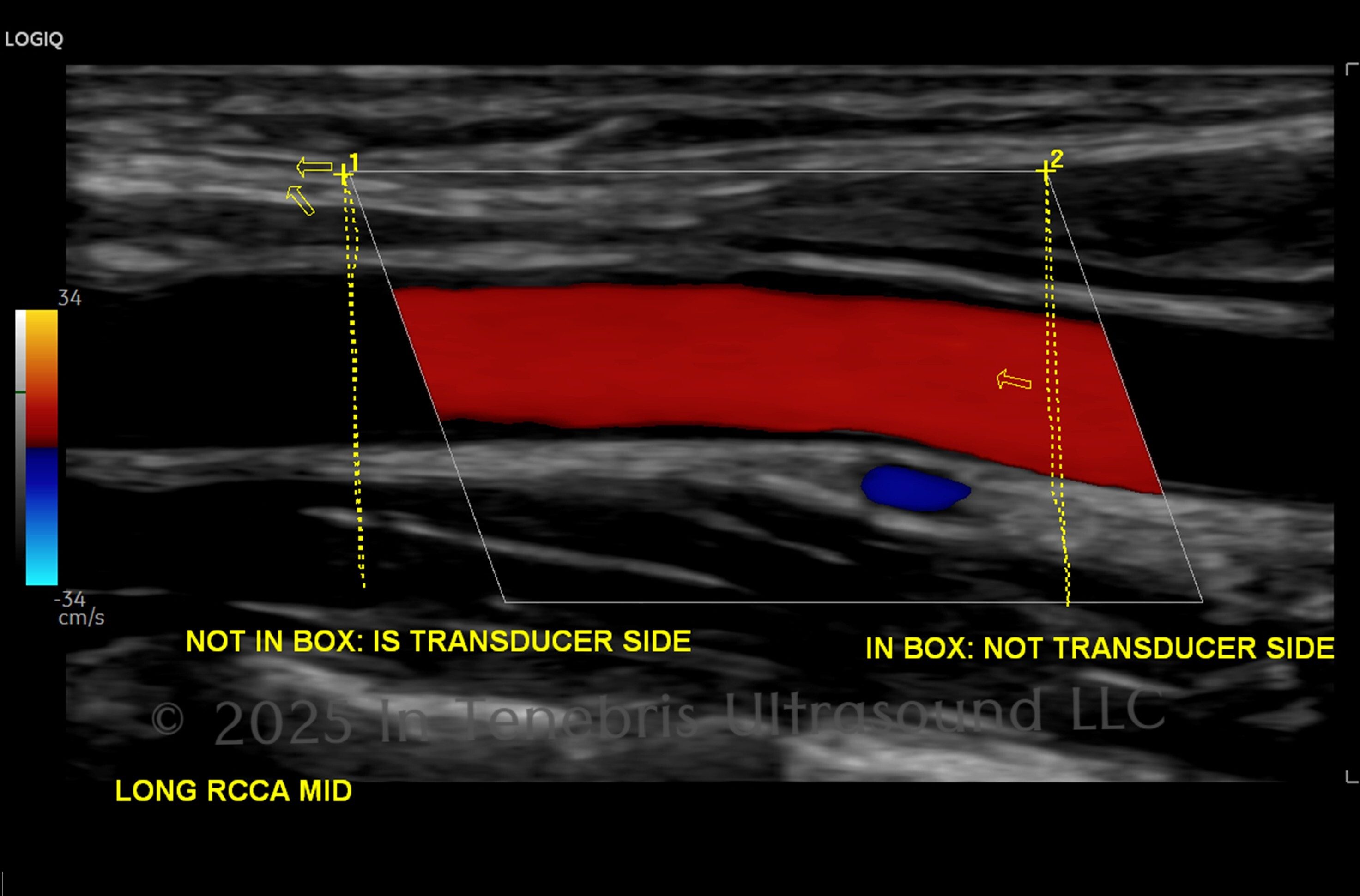
The color flow legend on the side of the screen shows what color is flowing toward the transducer (top color) and which is flowing away from the transducer (below the baseline). Which side is the transducer? The transducer side of the color box is the same side as the acute angle: you can draw a straight line down and stay outside of the box. You will often find pathology, collaterals, grafts, and unusual situations in which you have to determine blood flow direction, and this can get very confusing in the moment!
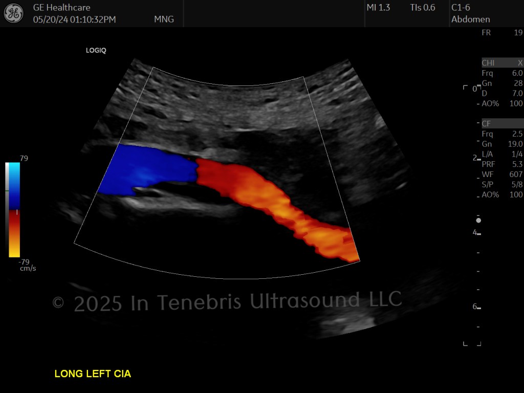
Curved Probe
The transducer is considered the line within the color box that is perpendicular to the vessel. You can window, angle, and rock the transducer so that the entire length of vessel is traveling toward or away from the transducer.
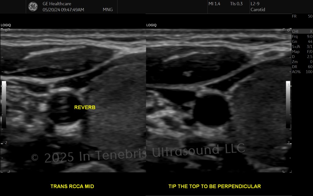
Guide to Clearing Out Vessels & Other Cystic Structures including Gallbladder & Bladder & Cysts
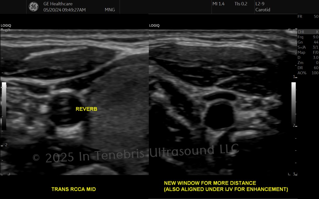
Artifact in vessels, the gallbladder, bladder, and even cysts looks like pathology: clot, plaque, solid lesions, etc. You have to clear the noise out, and no, TGCs are NOT the answer! The best optimization keeps the real echoes of the surrounding tissues, maintaining a consistent shade of gray from superficial to deep.
- Tip the top.
- More pressure (less distance) or less pressure (create more distance).
- New window: angle through more tissue or another structure such as internal jugular vein (IJV) or liver.
- Increase frequency.
- Increase speckle reduction imaging.
- Decrease overall power/gain.
- Increase compound imaging, frame averaging, line density.
- Create more distance with a standoff pad of gel (especially helpful for very superficial cysts or very skinny patients).
Guide to Palpables & Superficial Scans
I learned how to scan lumps when I was studying to scan breasts with precision. The best resource for studying for breast ultrasound, hands down, is Breast Ultrasound by Dr. Anthony Thomas Stavros (2004). This doctor is brilliant and has made tremendous contributions to the world of ultrasound. When you have to learn to scan hernias, obtain his article that is complete with cine clips (Stavros & Rapp, 2010). Even in researching morphology of arterial waveforms, his name as an expert investigator appears (Stavros et. al, 1992).
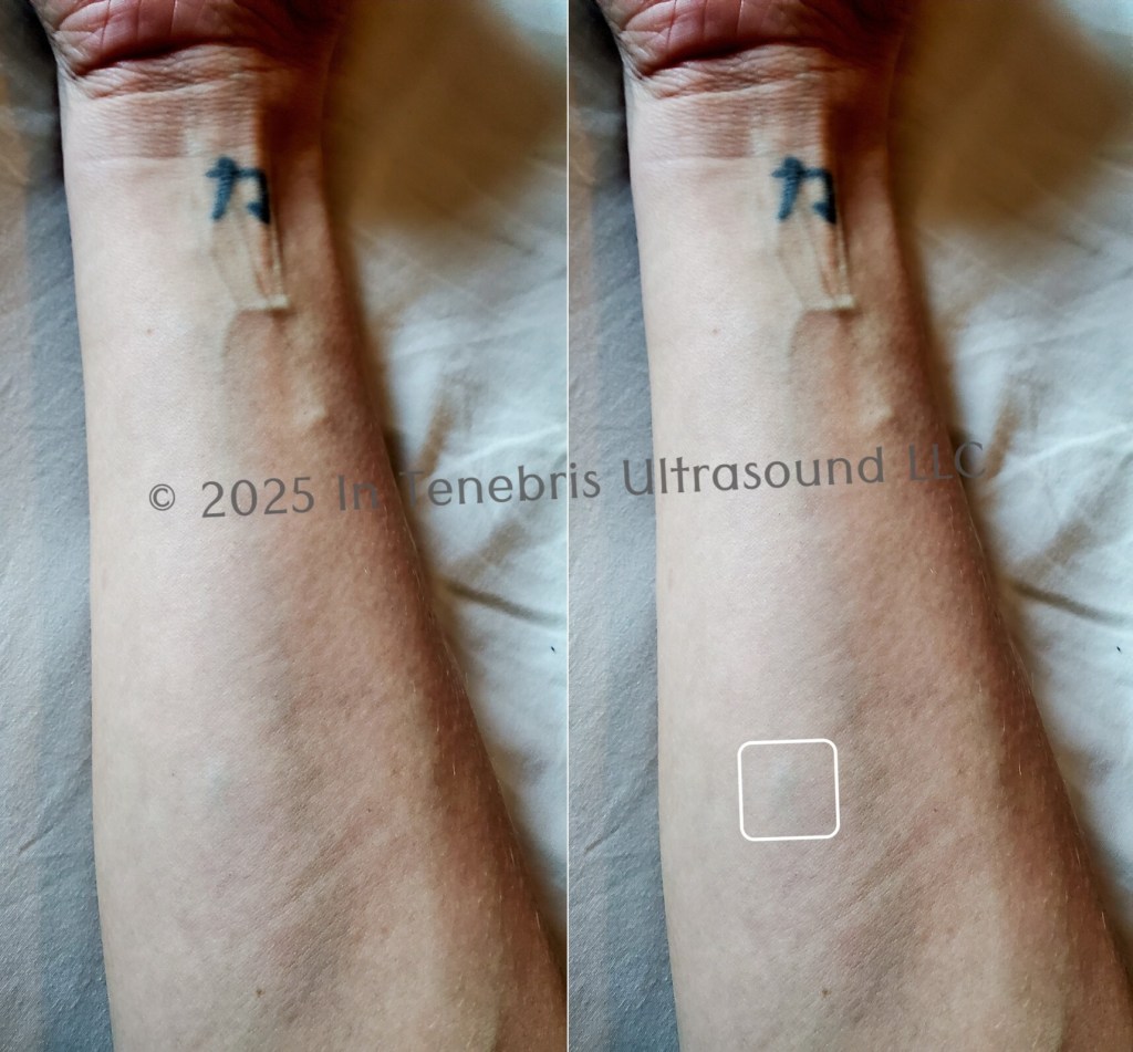
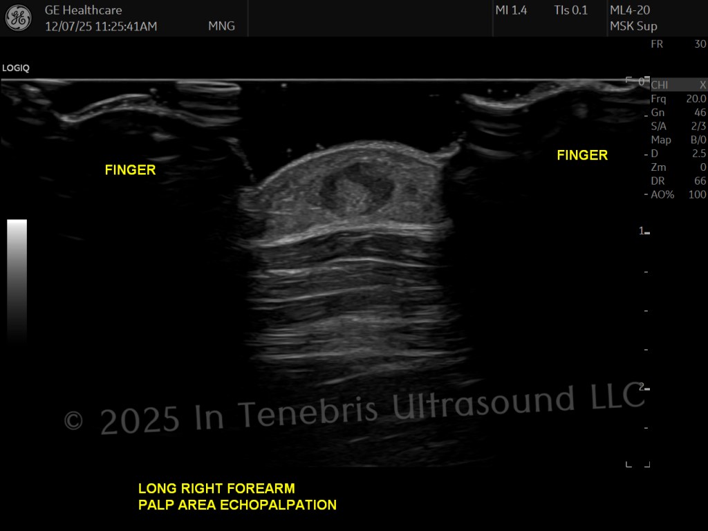
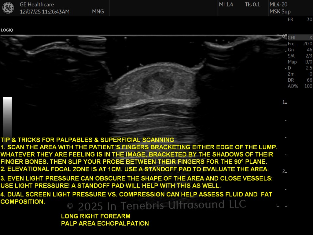
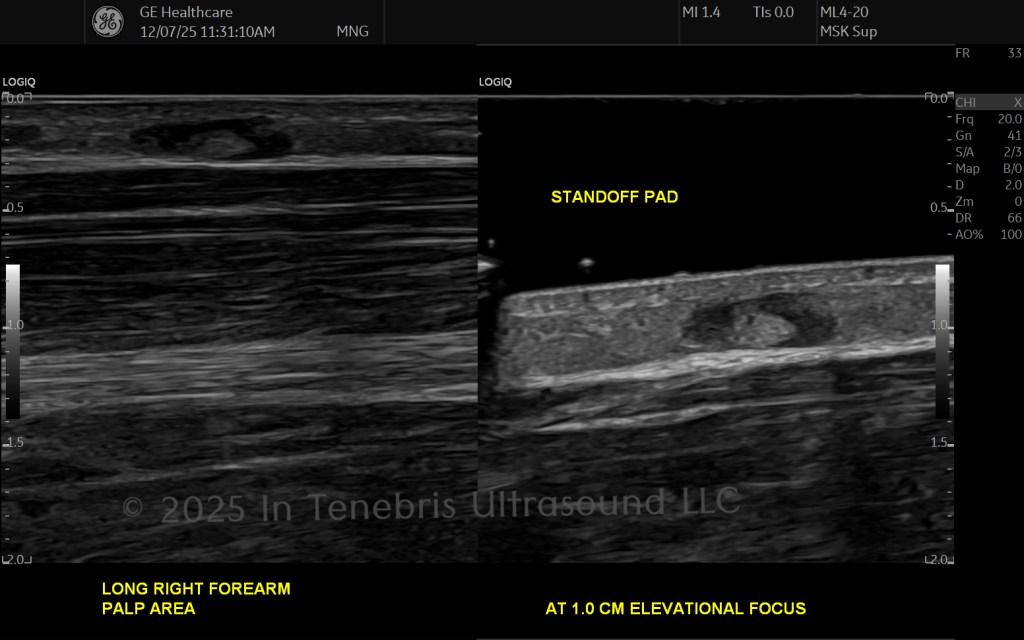
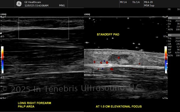
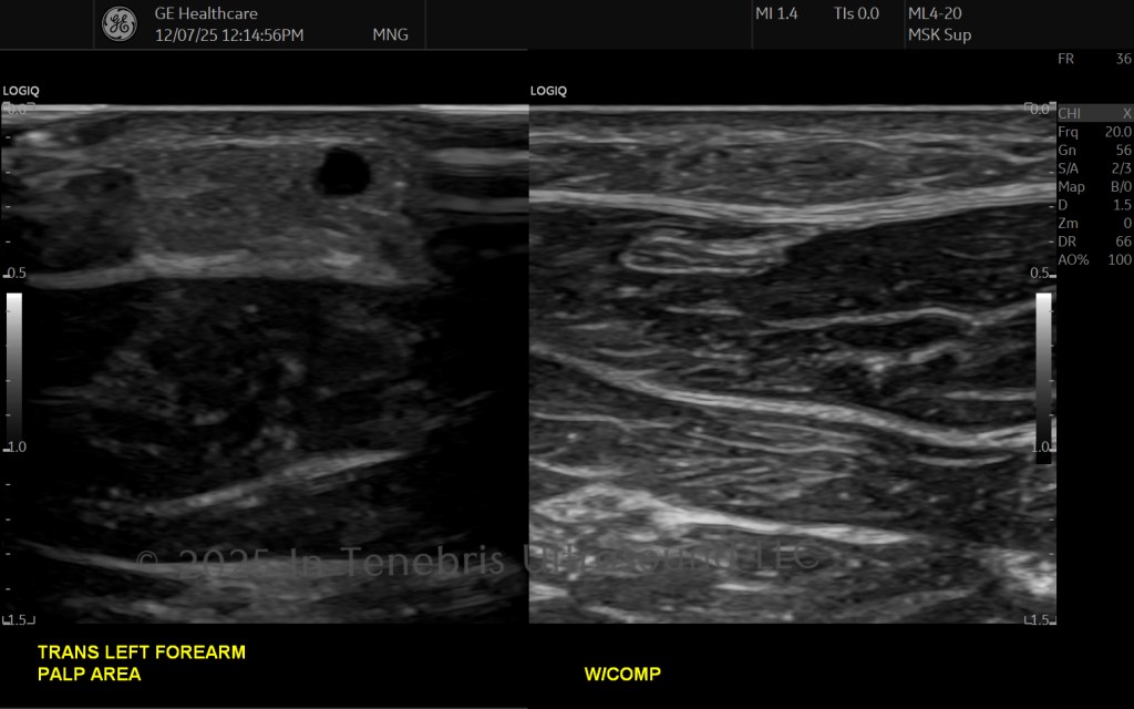
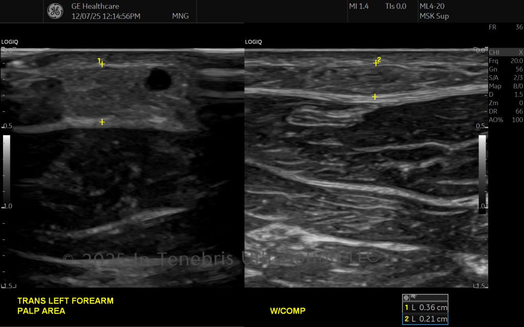
Echopalpation is a technique in which the sonographer scans (echo) the area while the area is being palpated either by the sonographer or the patient. This is an important step to utilize in scanning palpable areas—just because an area might be seen where a patient says they feel an area does not mean that area is the area that they are feeling. Furthermore, oftentimes an area of normal anatomy can cause a prominent lump for a patient. Certain areas of adipose tissue like to be what I lovingly refer to as little divas. By using this technique, we can show the patient that what they are feeling is being shown on the screen. We can correlate normal anatomy with what they are feeling and reassure them that what is being felt is normal, though lumpy, tissue. Additionally, the fingers palpating the superficial lump create a base to rest the probe and a pocket where we can fill the area with gel, creating a steady standoff pad. This is important to localize the area at the elevational focus which is typically 1.0 cm deep. The skill to use this technique takes practice but is an invaluable tool. Although this is a widely used technique, I personally learned the skill from Dr. Stavros’s book and feel indebted to him for the guidance (2004).
Physics Practice Problems (coming soon)
From simple wave equations to complex Doppler calculations, these posts present worked examples and exam-style challenges to build fluency and confidence.

Reference Formula Hub (coming soon)
This area compiles core formulas, clear definitions, and labeled diagrams so students can quickly locate and review essential ultrasound concepts.
